8U0Q | pdb_00008u0q

Co-crystal structure of optimized analog TDI-13537 provided new insights into the potency determinants of the sulfonamide inhibitor series


Experimental Data Snapshot

  • Method: X-RAY DIFFRACTION
  • Resolution: 1.69 Å
  • R-Value Free: 
    0.208 (Depositor), 0.199 (DCC) 
  • R-Value Work: 
    0.171 (Depositor) 
  • R-Value Observed: 
    0.173 (Depositor) 

Starting Model: experimental
View more details

wwPDB Validation 3D Report Full Report

Validation slider image for 8U0Q

Ligand Structure Quality Assessment 


This is version 1.1 of the entry. See complete history

Literature

Shape-Based Virtual Screening of a Billion-Compound Library Identifies Mycobacterial Lipoamide Dehydrogenase Inhibitors.

Michino, M.Beautrait, A.Boyles, N.A.Nadupalli, A.Dementiev, A.Sun, S.Ginn, J.Baxt, L.Suto, R.Bryk, R.Jerome, S.V.Huggins, D.J.Vendome, J.

(2023) ACS Bio Med Chem Au 3: 507-515

  • DOI: https://doi.org/10.1021/acsbiomedchemau.3c00046
  • Primary Citation Related Structures: 
    8U0Q

  • PubMed Abstract: 

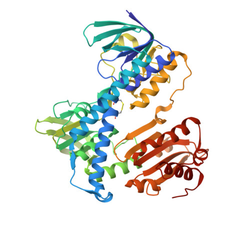
    Lpd (lipoamide dehydrogenase) in Mycobacterium tuberculosis (Mtb) is required for virulence and is a genetically validated tuberculosis (TB) target. Numerous screens have been performed over the last decade, yet only two inhibitor series have been identified. Recent advances in large-scale virtual screening methods combined with make-on-demand compound libraries have shown the potential for finding novel hits. In this study, the Enamine REAL library consisting of ∼1.12 billion compounds was efficiently screened using the GPU Shape screen method against Mtb Lpd to find additional chemical matter that would expand on the known sulfonamide inhibitor series. We identified six new inhibitors with IC 50 in the range of 5-100 μM. While these compounds remained chemically close to the already known sulfonamide series inhibitors, some diversity was found in the cores of the hits. The two most potent hits were further validated by one-step potency optimization to submicromolar levels. The co-crystal structure of optimized analogue TDI-13537 provided new insights into the potency determinants of the series.


  • Organizational Affiliation
    • Sanders Tri-Institutional Therapeutics Discovery Institute, 1230 York Avenue, Box 122, New York, New York 10065, United States.

Macromolecule Content 

  • Total Structure Weight: 104.61 kDa 
  • Atom Count: 7,887 
  • Modeled Residue Count: 928 
  • Deposited Residue Count: 932 
  • Unique protein chains: 1

Macromolecules

Find similar proteins by:|  3D Structure
Entity ID: 1
MoleculeChains  Sequence LengthOrganismDetailsImage
Dihydrolipoyl dehydrogenase
A, B
466Mycobacterium tuberculosisMutation(s): 0 
Gene Names: lpdClpdRv0462MTV038.06
EC: 1.8.1.4
UniProt
Find proteins for P9WHH9 (Mycobacterium tuberculosis (strain ATCC 25618 / H37Rv))
Explore P9WHH9 
Go to UniProtKB:  P9WHH9
Entity Groups
Sequence Clusters30% Identity50% Identity70% Identity90% Identity95% Identity100% Identity
UniProt GroupP9WHH9
Sequence Annotations
Expand
Reference Sequence

Small Molecules

Ligands 3 Unique
IDChains Name / Formula / InChI Key2D Diagram3D Interactions
FAD

Query on FAD


Download:Ideal Coordinates CCD File
BA [auth B],
C [auth A]
FLAVIN-ADENINE DINUCLEOTIDE
C27 H33 N9 O15 P2
VWWQXMAJTJZDQX-UYBVJOGSSA-N
U4I

Query on U4I


Download:Ideal Coordinates CCD File
D [auth A],
Y [auth A]
N-(3-acetamidophenyl)-N~2~-[3-(difluoromethyl)-5-methylbenzene-1-sulfonyl]-N~2~-methylglycinamide
C19 H21 F2 N3 O4 S
MYNNHYIXRANCAX-UHFFFAOYSA-N
GOL

Query on GOL


Download:Ideal Coordinates CCD File
AA [auth B]
CA [auth B]
DA [auth B]
E [auth A]
EA [auth B]
AA [auth B],
CA [auth B],
DA [auth B],
E [auth A],
EA [auth B],
F [auth A],
FA [auth B],
G [auth A],
GA [auth B],
H [auth A],
HA [auth B],
I [auth A],
IA [auth B],
J [auth A],
JA [auth B],
K [auth A],
KA [auth B],
L [auth A],
LA [auth B],
M [auth A],
MA [auth B],
N [auth A],
NA [auth B],
O [auth A],
OA [auth B],
P [auth A],
PA [auth B],
Q [auth A],
R [auth A],
S [auth A],
T [auth A],
U [auth A],
V [auth A],
W [auth A],
X [auth A],
Z [auth B]
GLYCEROL
C3 H8 O3
PEDCQBHIVMGVHV-UHFFFAOYSA-N

Experimental Data & Validation

Experimental Data

  • Method: X-RAY DIFFRACTION
  • Resolution: 1.69 Å
  • R-Value Free:  0.208 (Depositor), 0.199 (DCC) 
  • R-Value Work:  0.171 (Depositor) 
  • R-Value Observed: 0.173 (Depositor) 
Space Group: P 21 21 21
Unit Cell:
Length ( Å )Angle ( ˚ )
a = 83.335α = 90
b = 97.928β = 90
c = 123.806γ = 90
Software Package:
Software NamePurpose
PHENIXrefinement
XDSdata reduction
XDSdata scaling
PHASERphasing

Structure Validation

View Full Validation Report


Ligand Structure Quality Assessment 


Entry History 

& Funding Information

Deposition Data


Funding OrganizationLocationGrant Number
Not funded--

Revision History  (Full details and data files)

  • Version 1.0: 2024-01-03
    Type: Initial release
  • Version 1.1: 2024-11-13
    Changes: Structure summary