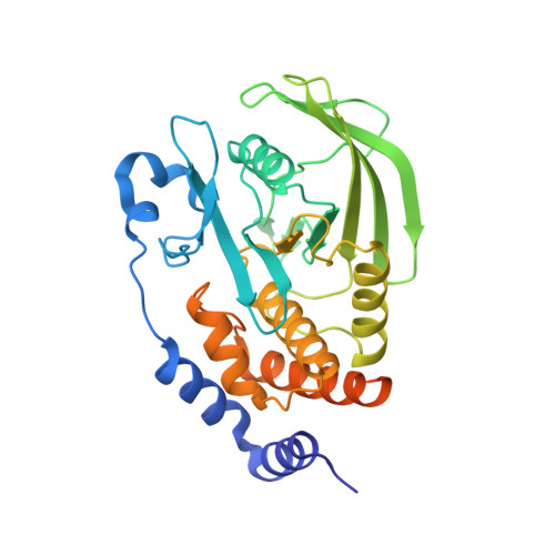
8U0H

Crystal structure of PTPN2 with a PROTAC


Experimental Data Snapshot

  • Method: X-RAY DIFFRACTION
  • Resolution: 1.93 Å
  • R-Value Free: 0.192 
  • R-Value Work: 0.173 
  • R-Value Observed: 0.173 

Starting Model: experimental
View more details

wwPDB Validation   3D Report Full Report


Ligand Structure Quality Assessment 


This is version 1.0 of the entry. See complete history


Literature

Mechanistic insights into a heterobifunctional degrader-induced PTPN2/N1 complex.

Hao, Q.Rathinaswamy, M.K.Klinge, K.L.Bratkowski, M.Mafi, A.Baumgartner, C.K.Hamel, K.M.Veits, G.K.Jain, R.Catalano, C.Fitzgerald, M.Hird, A.W.Park, E.Vora, H.U.Henderson, J.A.Longenecker, K.Hutchins, C.W.Qiu, W.Scapin, G.Sun, Q.Stoll, V.S.Sun, C.Li, P.Eaton, D.Stokoe, D.Fisher, S.L.Nasveschuk, C.G.Paddock, M.Kort, M.E.

(2024) Commun Chem 7: 183-183

  • DOI: https://doi.org/10.1038/s42004-024-01263-7
  • Primary Citation of Related Structures:  
    8U0H, 8UH6

  • PubMed Abstract: 

    PTPN2 (protein tyrosine phosphatase non-receptor type 2, or TC-PTP) and PTPN1 are attractive immuno-oncology targets, with the deletion of Ptpn1 and Ptpn2 improving response to immunotherapy in disease models. Targeted protein degradation has emerged as a promising approach to drug challenging targets including phosphatases. We developed potent PTPN2/N1 dual heterobifunctional degraders (Cmpd-1 and Cmpd-2) which facilitate efficient complex assembly with E3 ubiquitin ligase CRL4 CRBN , and mediate potent PTPN2/N1 degradation in cells and mice. To provide mechanistic insights into the cooperative complex formation introduced by degraders, we employed a combination of structural approaches. Our crystal structure reveals how PTPN2 is recognized by the tri-substituted thiophene moiety of the degrader. We further determined a high-resolution structure of DDB1-CRBN/Cmpd-1/PTPN2 using single-particle cryo-electron microscopy (cryo-EM). This structure reveals that the degrader induces proximity between CRBN and PTPN2, albeit the large conformational heterogeneity of this ternary complex. The molecular dynamic (MD)-simulations constructed based on the cryo-EM structure exhibited a large rigid body movement of PTPN2 and illustrated the dynamic interactions between PTPN2 and CRBN. Together, our study demonstrates the development of PTPN2/N1 heterobifunctional degraders with potential applications in cancer immunotherapy. Furthermore, the developed structural workflow could help to understand the dynamic nature of degrader-induced cooperative ternary complexes.


  • Organizational Affiliation

    Calico Life Sciences LLC, South San Francisco, CA, 94080, USA. qhao@calicolabs.com.


Macromolecules
Find similar proteins by:  (by identity cutoff)  |  3D Structure
Entity ID: 1
MoleculeChains Sequence LengthOrganismDetailsImage
PTPN2
A, B
324Homo sapiensMutation(s): 0 
Gene Names: PTPN2PTPT
EC: 3.1.3.48
UniProt & NIH Common Fund Data Resources
Find proteins for P17706 (Homo sapiens)
Explore P17706 
Go to UniProtKB:  P17706
PHAROS:  P17706
GTEx:  ENSG00000175354 
Entity Groups  
Sequence Clusters30% Identity50% Identity70% Identity90% Identity95% Identity100% Identity
UniProt GroupP17706
Sequence Annotations
Expand
  • Reference Sequence
Small Molecules
Ligands 3 Unique
IDChains Name / Formula / InChI Key2D Diagram3D Interactions
UB0 (Subject of Investigation/LOI)
Query on UB0

Download Ideal Coordinates CCD File 
C [auth A],
F [auth B]
(5P)-3-(carboxymethoxy)-4-chloro-5-(3-{[(4S)-1-({3-[2-(4-{3-[(3R)-2,6-dioxopiperidin-3-yl]-2-oxo-2,3-dihydro-1,3-benzoxazol-6-yl}piperidin-1-yl)acetamido]phenyl}methanesulfonyl)-2,2-dimethylpiperidin-4-yl]amino}phenyl)thiophene-2-carboxylic acid
C46 H49 Cl N6 O12 S2
XIVCTXQEWAGEQY-UZNNEEJFSA-N
ACT
Query on ACT

Download Ideal Coordinates CCD File 
D [auth A],
G [auth B]
ACETATE ION
C2 H3 O2
QTBSBXVTEAMEQO-UHFFFAOYSA-M
NA
Query on NA

Download Ideal Coordinates CCD File 
E [auth A]SODIUM ION
Na
FKNQFGJONOIPTF-UHFFFAOYSA-N
Experimental Data & Validation

Experimental Data

  • Method: X-RAY DIFFRACTION
  • Resolution: 1.93 Å
  • R-Value Free: 0.192 
  • R-Value Work: 0.173 
  • R-Value Observed: 0.173 
  • Space Group: C 2 2 21
Unit Cell:
Length ( Å )Angle ( ˚ )
a = 77.18α = 90
b = 126.51β = 90
c = 150.21γ = 90
Software Package:
Software NamePurpose
PHENIXrefinement
autoPROCdata reduction
XSCALEdata scaling
MOLREPphasing

Structure Validation

View Full Validation Report


Ligand Structure Quality Assessment 


Entry History & Funding Information

Deposition Data


Funding OrganizationLocationGrant Number
Not funded--

Revision History  (Full details and data files)

  • Version 1.0: 2024-09-04
    Type: Initial release