8U02 | pdb_00008u02

CryoEM structure of D2 dopamine receptor in complex with GoA KE mutant and dopamine


Experimental Data Snapshot

  • Method: ELECTRON MICROSCOPY
  • Resolution: 3.28 Å
  • Aggregation State: PARTICLE 
  • Reconstruction Method: SINGLE PARTICLE 

wwPDB Validation 3D Report Full Report

Validation slider image for 8U02

This is version 1.2 of the entry. See complete history. 

Literature

A neurodevelopmental disorder mutation locks G proteins in the transitory pre-activated state.

Knight, K.M., Krumm, B.E., Kapolka, N.J., Ludlam, W.G., Cui, M., Mani, S., Prytkova, I., Obarow, E.G., Lefevre, T.J., Wei, W., Ma, N., Huang, X.P., Fay, J.F., Vaidehi, N., Smrcka, A.V., Slesinger, P.A., Logothetis, D.E., Martemyanov, K.A., Roth, B.L., Dohlman, H.G.

(2024) Nat Commun 15: 6643-6643

  • DOI: https://doi.org/10.1038/s41467-024-50964-z
  • Primary Citation Related Structures: 
    8TZQ, 8U02

  • PubMed Abstract: 

    Many neurotransmitter receptors activate G proteins through exchange of GDP for GTP. The intermediate nucleotide-free state has eluded characterization, due largely to its inherent instability. Here we characterize a G protein variant associated with a rare neurological disorder in humans. Gα o K46E has a charge reversal that clashes with the phosphate groups of GDP and GTP. As anticipated, the purified protein binds poorly to guanine nucleotides yet retains wild-type affinity for G protein βγ subunits. In cells with physiological concentrations of nucleotide, Gα o K46E forms a stable complex with receptors and Gβγ, impeding effector activation. Further, we demonstrate that the mutant can be easily purified in complex with dopamine-bound D2 receptors, and use cryo-electron microscopy to determine the structure, including both domains of Gα o , without nucleotide or stabilizing nanobodies. These findings reveal the molecular basis for the first committed step of G protein activation, establish a mechanistic basis for a neurological disorder, provide a simplified strategy to determine receptor-G protein structures, and a method to detect high affinity agonist binding in cells.


  • Organizational Affiliation: 
    • Department of Pharmacology, University of North Carolina at Chapel Hill, Chapel Hill, NC, USA.

Macromolecule Content 

  • Total Structure Weight: 138.22 kDa 
  • Atom Count: 7,774 
  • Modeled Residue Count: 1,013 
  • Deposited Residue Count: 1,226 
  • Unique protein chains: 4

Macromolecules

Find similar proteins by:|  3D Structure
Entity ID: 1
MoleculeChains  Sequence LengthOrganismDetailsImage
D(2) dopamine receptorA [auth R]443Homo sapiensMutation(s): 0 
Gene Names: DRD2
Membrane Entity: Yes 
UniProt & NIH Common Fund Data Resources
Find proteins for P14416 (Homo sapiens)
Explore P14416 
Go to UniProtKB:  P14416
PHAROS:  P14416
GTEx:  ENSG00000149295 
Entity Groups
Sequence Clusters30% Identity50% Identity70% Identity90% Identity95% Identity100% Identity
UniProt GroupP14416
Sequence Annotations
Expand
Reference Sequence
Find similar proteins by:|  3D Structure
Entity ID: 2
MoleculeChains  Sequence LengthOrganismDetailsImage
Guanine nucleotide-binding protein G(o) subunit alpha354Homo sapiensMutation(s): 1 
Gene Names: GNAO1
EC: 3.6.5
Membrane Entity: Yes 
UniProt & NIH Common Fund Data Resources
Find proteins for P09471 (Homo sapiens)
Explore P09471 
Go to UniProtKB:  P09471
PHAROS:  P09471
GTEx:  ENSG00000087258 
Entity Groups
Sequence Clusters30% Identity50% Identity70% Identity90% Identity95% Identity100% Identity
UniProt GroupP09471
Sequence Annotations
Expand
Reference Sequence
Find similar proteins by:|  3D Structure
Entity ID: 3
MoleculeChains  Sequence LengthOrganismDetailsImage
Guanine nucleotide-binding protein G(I)/G(S)/G(T) subunit beta-1C [auth A]358Homo sapiensMutation(s): 0 
Gene Names: GNB1
UniProt & NIH Common Fund Data Resources
Find proteins for P62873 (Homo sapiens)
Explore P62873 
Go to UniProtKB:  P62873
PHAROS:  P62873
GTEx:  ENSG00000078369 
Entity Groups
Sequence Clusters30% Identity50% Identity70% Identity90% Identity95% Identity100% Identity
UniProt GroupP62873
Sequence Annotations
Expand
Reference Sequence
Find similar proteins by:|  3D Structure
Entity ID: 4
MoleculeChains  Sequence LengthOrganismDetailsImage
Guanine nucleotide-binding protein G(I)/G(S)/G(O) subunit gamma-2D [auth C]71Homo sapiensMutation(s): 0 
Gene Names: GNG2
Membrane Entity: Yes 
UniProt & NIH Common Fund Data Resources
Find proteins for P59768 (Homo sapiens)
Explore P59768 
Go to UniProtKB:  P59768
PHAROS:  P59768
GTEx:  ENSG00000186469 
Entity Groups
Sequence Clusters30% Identity50% Identity70% Identity90% Identity95% Identity100% Identity
UniProt GroupP59768
Sequence Annotations
Expand
Reference Sequence

Small Molecules

Ligands 1 Unique
IDChains Name / Formula / InChI Key2D Diagram3D Interactions
LDP
(Subject of Investigation/LOI)

Query on LDP


Download:Ideal Coordinates CCD File
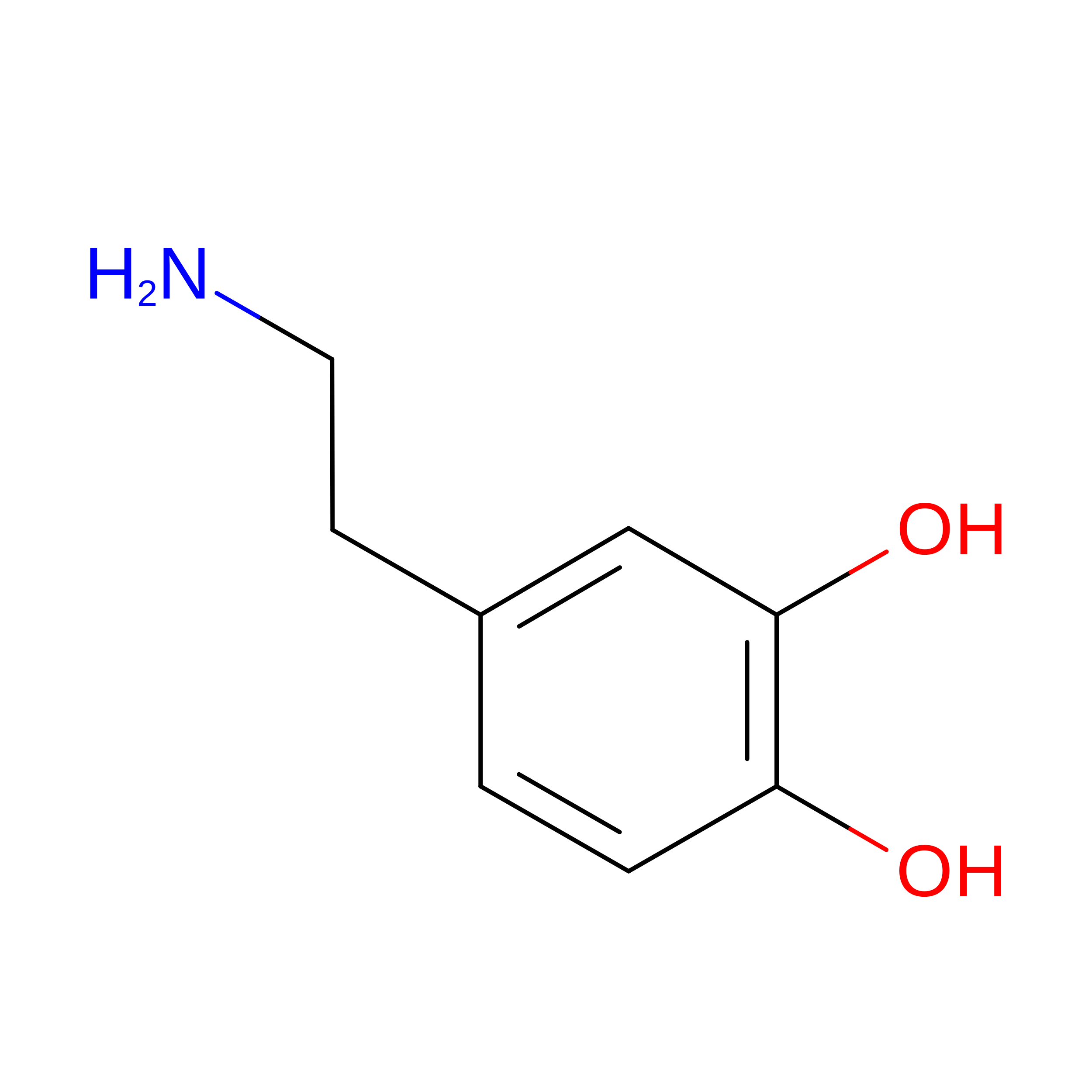
E [auth R]L-DOPAMINE
C8 H11 N O2
VYFYYTLLBUKUHU-UHFFFAOYSA-N
Binding Affinity Annotations 
IDSourceBinding Affinity
LDP BindingDB:  8U02 Ki: min: 1.8, max: 1.50e+4 (nM) from 75 assay(s)
Kd: min: 6200, max: 2.67e+5 (nM) from 5 assay(s)
IC50: 0 (nM) from 1 assay(s)
EC50: min: 0.63, max: 3460 (nM) from 39 assay(s)

Experimental Data & Validation

Experimental Data

  • Method: ELECTRON MICROSCOPY
  • Resolution: 3.28 Å
  • Aggregation State: PARTICLE 
  • Reconstruction Method: SINGLE PARTICLE 
EM Software:
TaskSoftware PackageVersion
MODEL REFINEMENTPHENIX

Structure Validation

View Full Validation Report


Entry History 

& Funding Information

Deposition Data


Funding OrganizationLocationGrant Number
National Institutes of Health/National Institute of Mental Health (NIH/NIMH)United StatesMH112205

Revision History  (Full details and data files)

  • Version 1.0: 2024-08-21
    Type: Initial release
  • Version 1.1: 2024-11-20
    Changes: Data collection, Structure summary
  • Version 1.2: 2025-05-28
    Changes: Data collection