8TUV

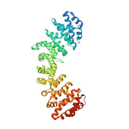
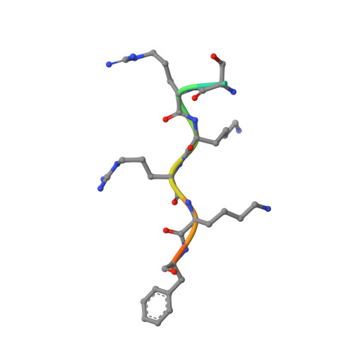
Merkel Cell Polyomavirus LTA NLSm bound to importin alpha 2


Experimental Data Snapshot

  • Method: X-RAY DIFFRACTION
  • Resolution: 2.30 Å
  • R-Value Free: 0.218 
  • R-Value Work: 0.191 
  • R-Value Observed: 0.193 

Starting Model: experimental
View more details

wwPDB Validation   3D Report Full Report


This is version 1.1 of the entry. See complete history


Literature

A functional and structural comparative analysis of large tumor antigens reveals evolution of different importin alpha-dependent nuclear localization signals.

Cross, E.M.Akbari, N.Ghassabian, H.Hoad, M.Pavan, S.Ariawan, D.Donnelly, C.M.Lavezzo, E.Petersen, G.F.Forwood, J.K.Alvisi, G.

(2024) Protein Sci 33: e4876-e4876

  • DOI: https://doi.org/10.1002/pro.4876
  • Primary Citation of Related Structures:  
    8Q8K, 8SUD, 8TUQ, 8TUR, 8TUS, 8TUT, 8TUU, 8TUV

  • PubMed Abstract: 

    Nucleocytoplasmic transport regulates the passage of proteins between the nucleus and cytoplasm. In the best characterized pathway, importin (IMP) α bridges cargoes bearing basic, classical nuclear localization signals (cNLSs) to IMPβ1, which mediates transport through the nuclear pore complex. IMPα recognizes three types of cNLSs via two binding sites: the major binding site accommodates monopartite cNLSs, the minor binding site recognizes atypical cNLSs, while bipartite cNLSs simultaneously interact with both major and minor sites. Despite the growing knowledge regarding IMPα-cNLS interactions, our understanding of the evolution of cNLSs is limited. We combined bioinformatic, biochemical, functional, and structural approaches to study this phenomenon, using polyomaviruses (PyVs) large tumor antigens (LTAs) as a model. We characterized functional cNLSs from all human (H)PyV LTAs, located between the LXCXE motif and origin binding domain. Surprisingly, the prototypical SV40 monopartite NLS is not well conserved; HPyV LTA NLSs are extremely heterogenous in terms of structural organization, IMPα isoform binding, and nuclear targeting abilities, thus influencing the nuclear accumulation properties of full-length proteins. While several LTAs possess bipartite cNLSs, merkel cell PyV contains a hybrid bipartite cNLS whose upstream stretch of basic amino acids can function as an atypical cNLS, specifically binding to the IMPα minor site upon deletion of the downstream amino acids after viral integration in the host genome. Therefore, duplication of a monopartite cNLS and subsequent accumulation of point mutations, optimizing interaction with distinct IMPα binding sites, led to the evolution of bipartite and atypical NLSs binding at the minor site.


  • Organizational Affiliation

    School of Dentistry and Medical Sciences, Charles Sturt University, Wagga Wagga, Australia.


Macromolecules
Find similar proteins by:  (by identity cutoff)  |  3D Structure
Entity ID: 1
MoleculeChains Sequence LengthOrganismDetailsImage
Importin subunit alpha-1510Mus musculusMutation(s): 0 
Gene Names: Kpna2Rch1
UniProt & NIH Common Fund Data Resources
Find proteins for P52293 (Mus musculus)
Explore P52293 
Go to UniProtKB:  P52293
IMPC:  MGI:103561
Entity Groups  
Sequence Clusters30% Identity50% Identity70% Identity90% Identity95% Identity100% Identity
UniProt GroupP52293
Sequence Annotations
Expand
  • Reference Sequence

Find similar proteins by:  Sequence   |   3D Structure  

Entity ID: 2
MoleculeChains Sequence LengthOrganismDetailsImage
SER-ARG-LYS-ARG-LYS-PHE11Merkel cell polyomavirusMutation(s): 0 
UniProt
Find proteins for T2HFI5 (Merkel cell polyomavirus)
Explore T2HFI5 
Go to UniProtKB:  T2HFI5
Entity Groups  
Sequence Clusters30% Identity50% Identity70% Identity90% Identity95% Identity100% Identity
UniProt GroupT2HFI5
Sequence Annotations
Expand
  • Reference Sequence
Find similar proteins by:  (by identity cutoff)  |  3D Structure
Entity ID: 3
MoleculeChains Sequence LengthOrganismDetailsImage
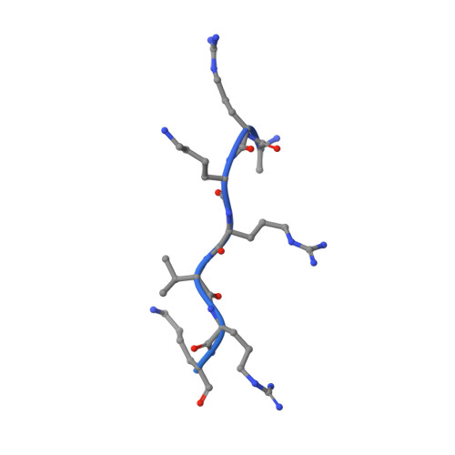
30S ribosomal protein S11129Escherichia coliMutation(s): 0 
UniProt
Find proteins for P0A7R9 (Escherichia coli (strain K12))
Explore P0A7R9 
Go to UniProtKB:  P0A7R9
Entity Groups  
Sequence Clusters30% Identity50% Identity70% Identity90% Identity95% Identity100% Identity
UniProt GroupP0A7R9
Sequence Annotations
Expand
  • Reference Sequence
Experimental Data & Validation

Experimental Data

  • Method: X-RAY DIFFRACTION
  • Resolution: 2.30 Å
  • R-Value Free: 0.218 
  • R-Value Work: 0.191 
  • R-Value Observed: 0.193 
  • Space Group: P 21 21 21
Unit Cell:
Length ( Å )Angle ( ˚ )
a = 77.273α = 90
b = 89.516β = 90
c = 97.221γ = 90
Software Package:
Software NamePurpose
PHENIXrefinement
XDSdata reduction
Cootmodel building
PHASERphasing
Aimlessdata scaling

Structure Validation

View Full Validation Report


Entry History & Funding Information

Deposition Data


Funding OrganizationLocationGrant Number
Not funded--

Revision History  (Full details and data files)

  • Version 1.0: 2024-02-07
    Type: Initial release
  • Version 1.1: 2024-02-14
    Changes: Database references