8TMT | pdb_00008tmt

Crystal structure of KPC-44 carbapenemase in complex with vaborbactam


Experimental Data Snapshot

  • Method: X-RAY DIFFRACTION
  • Resolution: 1.70 Å
  • R-Value Free: 
    0.201 (Depositor), 0.203 (DCC) 
  • R-Value Work: 
    0.161 (Depositor), 0.163 (DCC) 
  • R-Value Observed: 
    0.163 (Depositor) 

Starting Model: experimental
View more details

wwPDB Validation 3D Report Full Report

Validation slider image for 8TMT

Ligand Structure Quality Assessment 


This is version 1.2 of the entry. See complete history

Literature

Klebsiella pneumoniae carbapenemase variant 44 acquires ceftazidime-avibactam resistance by altering the conformation of active-site loops.

Sun, Z.Lin, H.Hu, L.Neetu, N.Sankaran, B.Wang, J.Prasad, B.V.V.Palzkill, T.

(2023) J Biological Chem 300: 105493-105493

  • DOI: https://doi.org/10.1016/j.jbc.2023.105493
  • Primary Citation Related Structures: 
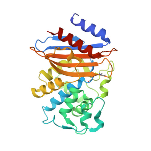
    8TJM, 8TMR, 8TMT, 8TN0

  • PubMed Abstract: 

    Klebsiella pneumoniae carbapenemase 2 (KPC-2) is an important source of drug resistance as it can hydrolyze and inactivate virtually all β-lactam antibiotics. KPC-2 is potently inhibited by avibactam via formation of a reversible carbamyl linkage of the inhibitor with the catalytic serine of the enzyme. However, the use of avibactam in combination with ceftazidime (CAZ-AVI) has led to the emergence of CAZ-AVI-resistant variants of KPC-2 in clinical settings. One such variant, KPC-44, bears a 15 amino acid duplication in one of the active-site loops (270-loop). Here, we show that the KPC-44 variant exhibits higher catalytic efficiency in hydrolyzing ceftazidime, lower efficiency toward imipenem and meropenem, and a similar efficiency in hydrolyzing ampicillin, than the WT KPC-2 enzyme. In addition, the KPC-44 variant enzyme exhibits 12-fold lower AVI carbamylation efficiency than the KPC-2 enzyme. An X-ray crystal structure of KPC-44 showed that the 15 amino acid duplication results in an extended and partially disordered 270-loop and also changes the conformation of the adjacent 240-loop, which in turn has altered interactions with the active-site omega loop. Furthermore, a structure of KPC-44 with avibactam revealed that formation of the covalent complex results in further disorder in the 270-loop, suggesting that rearrangement of the 270-loop of KPC-44 facilitates AVI carbamylation. These results suggest that the duplication of 15 amino acids in the KPC-44 enzyme leads to resistance to CAZ-AVI by modulating the stability and conformation of the 270-, 240-, and omega-loops.


  • Organizational Affiliation
    • Verna and Marrs McLean Department of Biochemistry and Molecular Pharmacology, Baylor College of Medicine, Houston, Texas, USA.

Macromolecule Content 

  • Total Structure Weight: 30.73 kDa 
  • Atom Count: 2,280 
  • Modeled Residue Count: 259 
  • Deposited Residue Count: 282 
  • Unique protein chains: 1

Macromolecules

Find similar proteins by:|  3D Structure
Entity ID: 1
MoleculeChains  Sequence LengthOrganismDetailsImage
beta-lactamase282Klebsiella pneumoniaeMutation(s): 0 
Gene Names: blaKPC
EC: 3.5.2.6
UniProt
Find proteins for A0A4Y5JTU1 (Klebsiella pneumoniae)
Explore A0A4Y5JTU1 
Go to UniProtKB:  A0A4Y5JTU1
Entity Groups
Sequence Clusters30% Identity50% Identity70% Identity90% Identity95% Identity100% Identity
UniProt GroupA0A4Y5JTU1
Sequence Annotations
Expand
Reference Sequence

Small Molecules

Ligands 6 Unique
IDChains Name / Formula / InChI Key2D Diagram3D Interactions
4D6
(Subject of Investigation/LOI)

Query on 4D6


Download:Ideal Coordinates CCD File
B [auth A]Vaborbactam
C12 H16 B N O5 S
IOOWNWLVCOUUEX-WPRPVWTQSA-N
SO4

Query on SO4


Download:Ideal Coordinates CCD File
H [auth A],
I [auth A]
SULFATE ION
O4 S
QAOWNCQODCNURD-UHFFFAOYSA-L
GOL

Query on GOL


Download:Ideal Coordinates CCD File
C [auth A]GLYCEROL
C3 H8 O3
PEDCQBHIVMGVHV-UHFFFAOYSA-N
EDO

Query on EDO


Download:Ideal Coordinates CCD File
E [auth A]1,2-ETHANEDIOL
C2 H6 O2
LYCAIKOWRPUZTN-UHFFFAOYSA-N
NA

Query on NA


Download:Ideal Coordinates CCD File
D [auth A],
G [auth A]
SODIUM ION
Na
FKNQFGJONOIPTF-UHFFFAOYSA-N
LI

Query on LI


Download:Ideal Coordinates CCD File
F [auth A]LITHIUM ION
Li
HBBGRARXTFLTSG-UHFFFAOYSA-N
Binding Affinity Annotations 
IDSourceBinding Affinity
4D6 BindingDB:  8TMT Ki: 56 (nM) from 1 assay(s)

Experimental Data & Validation

Experimental Data

  • Method: X-RAY DIFFRACTION
  • Resolution: 1.70 Å
  • R-Value Free:  0.201 (Depositor), 0.203 (DCC) 
  • R-Value Work:  0.161 (Depositor), 0.163 (DCC) 
  • R-Value Observed: 0.163 (Depositor) 
Space Group: P 32 2 1
Unit Cell:
Length ( Å )Angle ( ˚ )
a = 72.38α = 90
b = 72.38β = 90
c = 85.5γ = 120
Software Package:
Software NamePurpose
PHENIXrefinement
iMOSFLMdata reduction
SCALAdata scaling
PHASERphasing

Structure Validation

View Full Validation Report


Ligand Structure Quality Assessment 


Entry History 

& Funding Information

Deposition Data


Funding OrganizationLocationGrant Number
National Institutes of Health/National Institute Of Allergy and Infectious Diseases (NIH/NIAID)United StatesAI32956
Welch FoundationUnited StatesQ1279

Revision History  (Full details and data files)

  • Version 1.0: 2023-12-06
    Type: Initial release
  • Version 1.1: 2023-12-27
    Changes: Database references
  • Version 1.2: 2024-11-06
    Changes: Structure summary