


Structure-based functional analysis reveals multiple roles and widespread use of urea-binding proteins in nitrogen metabolism
Allert, M.J., Kumar, S., Wang, Y., Beese, L.S., Hellinga, H.W.To be published.
Experimental Data Snapshot
Starting Model: experimental
View more details
Entity ID: 1 | |||||
|---|---|---|---|---|---|
| Molecule | Chains | Sequence Length | Organism | Details | Image |
| Extracellular ligand-binding receptor | 393 | Caldicellulosiruptor saccharolyticus | Mutation(s): 0 Gene Names: Csac_2475 | 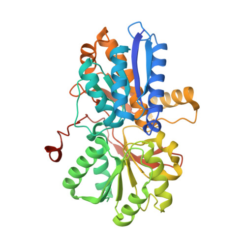 | |
UniProt | |||||
Entity Groups | |||||
| Sequence Clusters | 30% Identity50% Identity70% Identity90% Identity95% Identity100% Identity | ||||
| UniProt Group | A4XMB7 | ||||
Sequence AnnotationsExpand | |||||
Reference Sequence | |||||
| Ligands 2 Unique | |||||
|---|---|---|---|---|---|
| ID | Chains | Name / Formula / InChI Key | 2D Diagram | 3D Interactions | |
| BR Download:Ideal Coordinates CCD File | D [auth A] | BROMIDE ION Br CPELXLSAUQHCOX-UHFFFAOYSA-M |  | ||
| URE (Subject of Investigation/LOI) Download:Ideal Coordinates CCD File | C [auth A], E [auth B] | UREA C H4 N2 O XSQUKJJJFZCRTK-UHFFFAOYSA-N |  | ||
| Length ( Å ) | Angle ( ˚ ) |
|---|---|
| a = 79.235 | α = 90 |
| b = 91.67 | β = 90 |
| c = 96.546 | γ = 90 |
| Software Name | Purpose |
|---|---|
| PHENIX | refinement |
| XDS | data reduction |
| XDS | data scaling |
| PHASER | phasing |
| Funding Organization | Location | Grant Number |
|---|---|---|
| Becton-Dickinson and Company | United States | -- |


