Screening a knowledge-based library of low molecular weight compounds against the proline biosynthetic enzyme 1-pyrroline-5-carboxylate 1 (PYCR1).
Meeks, K.R., Bogner, A.N., Tanner, J.J.(2024) Protein Sci 33: e5072-e5072
- PubMed: 39133178 Search on PubMedSearch on PubMed Central
- DOI: https://doi.org/10.1002/pro.5072
- Primary Citation Related Structures:
8TD2, 8TD3, 8TD4, 8TD5, 8TD6, 8TD7, 8TD8, 8TD9, 8TDB, 8TDC, 8TDD, 8VRE, 8W0K - PubMed Abstract:
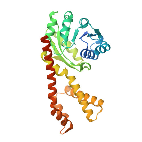
Δ 1 -pyrroline-5-carboxylate reductase isoform 1 (PYCR1) is the last enzyme of proline biosynthesis and catalyzes the NAD(P)H-dependent reduction of Δ 1 -pyrroline-5-carboxylate to L-proline. High PYCR1 gene expression is observed in many cancers and linked to poor patient outcomes and tumor aggressiveness. The knockdown of the PYCR1 gene or the inhibition of PYCR1 enzyme has been shown to inhibit tumorigenesis in cancer cells and animal models of cancer, motivating inhibitor discovery. We screened a library of 71 low molecular weight compounds (average MW of 131 Da) against PYCR1 using an enzyme activity assay. Hit compounds were validated with X-ray crystallography and kinetic assays to determine affinity parameters. The library was counter-screened against human Δ 1 -pyrroline-5-carboxylate reductase isoform 3 and proline dehydrogenase (PRODH) to assess specificity/promiscuity. Twelve PYCR1 and one PRODH inhibitor crystal structures were determined. Three compounds inhibit PYCR1 with competitive inhibition parameter of 100 μM or lower. Among these, (S)-tetrahydro-2H-pyran-2-carboxylic acid (70 μM) has higher affinity than the current best tool compound N-formyl-l-proline, is 30 times more specific for PYCR1 over human Δ 1 -pyrroline-5-carboxylate reductase isoform 3, and negligibly inhibits PRODH. Structure-affinity relationships suggest that hydrogen bonding of the heteroatom of this compound is important for binding to PYCR1. The structures of PYCR1 and PRODH complexed with 1-hydroxyethane-1-sulfonate demonstrate that the sulfonate group is a suitable replacement for the carboxylate anchor. This result suggests that the exploration of carboxylic acid isosteres may be a promising strategy for discovering new classes of PYCR1 and PRODH inhibitors. The structure of PYCR1 complexed with l-pipecolate and NADH supports the hypothesis that PYCR1 has an alternative function in lysine metabolism.
- Department of Biochemistry, University of Missouri, Columbia, Missouri, USA.
Organizational Affiliation: