Targeting the Structural Maturation Pathway of HIV-1 Reverse Transcriptase.
Kirby, T.W., Gabel, S.A., DeRose, E.F., Perera, L., Krahn, J.M., Pedersen, L.C., London, R.E.(2023) Biomolecules 13
- PubMed: 38002285 Search on PubMedSearch on PubMed Central
- DOI: https://doi.org/10.3390/biom13111603
- Primary Citation Related Structures:
8TCJ, 8TCK, 8TCL, 8TCM - PubMed Abstract:
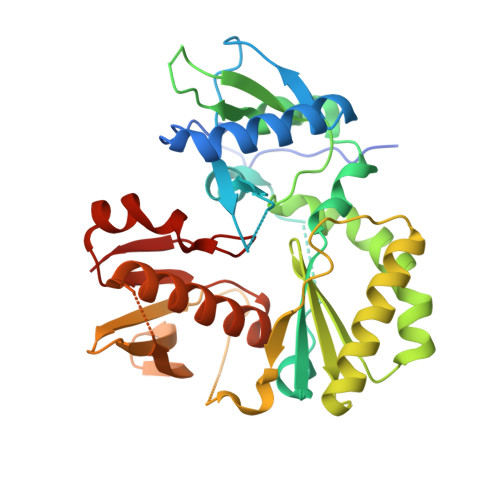
Formation of active HIV-1 reverse transcriptase (RT) proceeds via a structural maturation process that involves subdomain rearrangements and formation of an asymmetric p66/p66' homodimer. These studies were undertaken to evaluate whether the information about this maturation process can be used to identify small molecule ligands that retard or interfere with the steps involved. We utilized the isolated polymerase domain, p51, rather than p66, since the initial subdomain rearrangements are largely limited to this domain. Target sites at subdomain interfaces were identified and computational analysis used to obtain an initial set of ligands for screening. Chromatographic evaluations of the p51 homodimer/monomer ratio support the feasibility of this approach. Ligands that bind near the interfaces and a ligand that binds directly to a region of the fingers subdomain involved in subunit interface formation were identified, and the interactions were further characterized by NMR spectroscopy and X-ray crystallography. Although these ligands were found to reduce dimer formation, further efforts will be required to obtain ligands with higher binding affinity. In contrast with previous ligand identification studies performed on the RT heterodimer, subunit interface surfaces are solvent-accessible in the p51 and p66 monomers, making these constructs preferable for identification of ligands that directly interfere with dimerization.
- Genome Integrity and Structural Biology Laboratory, National Institute of Environmental Health Sciences, NIH, Research Triangle Park, Durham, NC 27709, USA.
Organizational Affiliation: