A Highly Ordered Nitroxide Side Chain for Distance Mapping and Monitoring Slow Structural Fluctuations in Proteins.
Chen, M., Kalai, T., Cascio, D., Bridges, M.D., Whitelegge, J.P., Elgeti, M., Hubbell, W.L.(2024) Appl Magn Reson 55: 251-277
- PubMed: 38357006 Search on PubMedSearch on PubMed Central
- DOI: https://doi.org/10.1007/s00723-023-01618-8
- Primary Citation Related Structures:
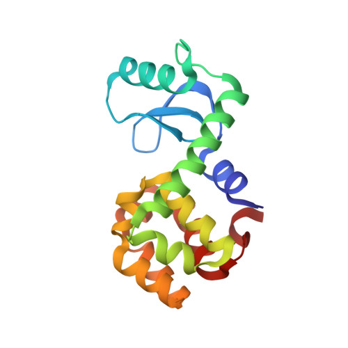
8TAT - PubMed Abstract:
Site-directed spin labeling electron paramagnetic resonance (SDSL-EPR) is an established tool for exploring protein structure and dynamics. Although nitroxide side chains attached to a single cysteine via a disulfide linkage are commonly employed in SDSL-EPR, their internal flexibility complicates applications to monitor slow internal motions in proteins and to structure determination by distance mapping. Moreover, the labile disulfide linkage prohibits the use of reducing agents often needed for protein stability. To enable the application of SDSL-EPR to the measurement of slow internal dynamics, new spin labels with hindered internal motion are desired. Here, we introduce a highly ordered nitroxide side chain, designated R9, attached at a single cysteine residue via a non-reducible thioether linkage. The reaction to introduce R9 is highly selective for solvent-exposed cysteine residues. Structures of R9 at two helical sites in T4 Lysozyme were determined by X-ray crystallography and the mobility in helical sequences was characterized by EPR spectral lineshape analysis, Saturation Transfer EPR, and Saturation Recovery EPR. In addition, interspin distance measurements between pairs of R9 residues are reported. Collectively, all data indicate that R9 will be useful for monitoring slow internal structural fluctuations, and applications to distance mapping via dipolar spectroscopy and relaxation enhancement methods are anticipated. The online version contains supplementary material available at 10.1007/s00723-023-01618-8.
- Jules Stein Eye Institute and Department of Chemistry and Biochemistry, University of California, Los Angeles, CA 90095 USA.
Organizational Affiliation: