8T2S

Structure of a group II intron ribonucleoprotein in the pre-branching (pre-1F) state


Experimental Data Snapshot

  • Method: ELECTRON MICROSCOPY
  • Resolution: 3.00 Å
  • Aggregation State: PARTICLE 
  • Reconstruction Method: SINGLE PARTICLE 

wwPDB Validation   3D Report Full Report


This is version 1.2 of the entry. See complete history


Literature

Structural insights into intron catalysis and dynamics during splicing.

Xu, L.Liu, T.Chung, K.Pyle, A.M.

(2023) Nature 624: 682-688

  • DOI: https://doi.org/10.1038/s41586-023-06746-6
  • Primary Citation of Related Structures:  
    8T2R, 8T2S, 8T2T

  • PubMed Abstract: 

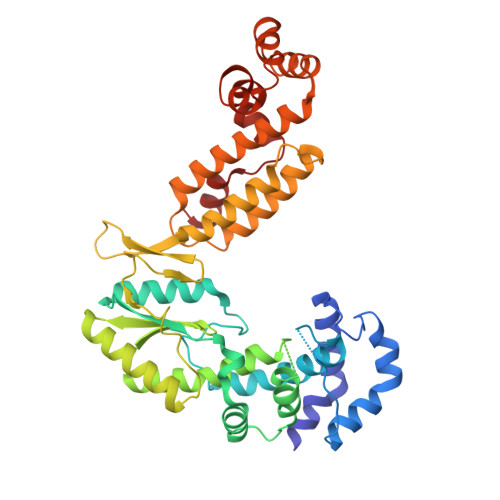
    The group II intron ribonucleoprotein is an archetypal splicing system with numerous mechanistic parallels to the spliceosome, including excision of lariat introns 1,2 . Despite the importance of branching in RNA metabolism, structural understanding of this process has remained elusive. Here we present a comprehensive analysis of three single-particle cryogenic electron microscopy structures captured along the splicing pathway. They reveal the network of molecular interactions that specifies the branchpoint adenosine and positions key functional groups to catalyse lariat formation and coordinate exon ligation. The structures also reveal conformational rearrangements of the branch helix and the mechanism of splice site exchange that facilitate the transition from branching to ligation. These findings shed light on the evolution of splicing and highlight the conservation of structural components, catalytic mechanism and dynamical strategies retained through time in premessenger RNA splicing machines.


  • Organizational Affiliation

    Howard Hughes Medical Institute, Chevy Chase, MD, USA. ling.xu@yale.edu.


Macromolecules

Find similar proteins by:  (by identity cutoff)  |  3D Structure
Entity ID: 1
MoleculeChains Sequence LengthOrganismDetailsImage
Group II intron reverse transcriptase/maturaseA [auth D]427Agathobacter rectalisMutation(s): 0 
Gene Names: ltrA_2ltrAERS852417_00966FYL37_05080
EC: 2.7.7.49
UniProt
Find proteins for A0A173ZME3 (Agathobacter rectalis)
Explore A0A173ZME3 
Go to UniProtKB:  A0A173ZME3
Entity Groups  
Sequence Clusters30% Identity50% Identity70% Identity90% Identity95% Identity100% Identity
UniProt GroupA0A173ZME3
Sequence Annotations
Expand
  • Reference Sequence
Find similar nucleic acids by:  (by identity cutoff)  |  3D Structure
Entity ID: 2
MoleculeChains LengthOrganismImage
RNA (551-MER)652Agathobacter rectalis
Sequence Annotations
Expand
  • Reference Sequence
Small Molecules
Ligands 2 Unique
IDChains Name / Formula / InChI Key2D Diagram3D Interactions
CA (Subject of Investigation/LOI)
Query on CA

Download Ideal Coordinates CCD File 
C [auth B]
D [auth B]
E [auth B]
F [auth B]
G [auth B]
C [auth B],
D [auth B],
E [auth B],
F [auth B],
G [auth B],
H [auth B],
I [auth B],
J [auth B],
K [auth B],
L [auth B],
M [auth B]
CALCIUM ION
Ca
BHPQYMZQTOCNFJ-UHFFFAOYSA-N
NH4 (Subject of Investigation/LOI)
Query on NH4

Download Ideal Coordinates CCD File 
N [auth B],
O [auth B]
AMMONIUM ION
H4 N
QGZKDVFQNNGYKY-UHFFFAOYSA-O
Experimental Data & Validation

Experimental Data

  • Method: ELECTRON MICROSCOPY
  • Resolution: 3.00 Å
  • Aggregation State: PARTICLE 
  • Reconstruction Method: SINGLE PARTICLE 
EM Software:
TaskSoftware PackageVersion
MODEL REFINEMENTPHENIX1.20.1_4487:

Structure Validation

View Full Validation Report


Entry History & Funding Information

Deposition Data


Funding OrganizationLocationGrant Number
Howard Hughes Medical Institute (HHMI)United States--

Revision History  (Full details and data files)

  • Version 1.0: 2023-11-22
    Type: Initial release
  • Version 1.1: 2023-12-06
    Changes: Database references
  • Version 1.2: 2024-01-03
    Changes: Database references