8SYK | pdb_00008syk

Crystal structure of RNA device 43 truncation mutant 3 (U100C), holo state


Experimental Data Snapshot

  • Method: X-RAY DIFFRACTION
  • Resolution: 3.06 Å
  • R-Value Free: 
    0.257 (Depositor), 0.258 (DCC) 
  • R-Value Work: 
    0.225 (Depositor), 0.225 (DCC) 
  • R-Value Observed: 
    0.227 (Depositor) 

Starting Model: experimental
View more details

wwPDB Validation 3D Report Full Report

Validation slider image for 8SYK

Ligand Structure Quality Assessment 


This is version 1.1 of the entry. See complete history

Literature

Structural investigation of an RNA device that regulates PD-1 expression in mammalian cells.

Stagno, J.R.Deme, J.C.Dwivedi, V.Lee, Y.T.Lee, H.K.Yu, P.Chen, S.Y.Fan, L.Degenhardt, M.F.S.Chari, R.Young, H.A.Lea, S.M.Wang, Y.X.

(2025) Nucleic Acids Res 53

  • DOI: https://doi.org/10.1093/nar/gkaf156
  • Primary Citation Related Structures: 
    8SYK, 8T5O, 8TKJ, 8TKK

  • PubMed Abstract: 

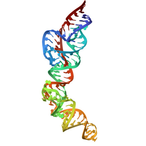
    Synthetic RNA devices are engineered to control gene expression and offer great potential in both biotechnology and clinical applications. Here, we present multidisciplinary structural and biochemical data for a tetracycline (Tc)-responsive RNA device (D43) in both ligand-free and bound states, providing a structure-dynamical basis for signal transmission. Activation of self-cleavage is achieved via ligand-induced conformational and dynamical changes that stabilize the elongated bridging helix harboring the communication module, which drives proper coordination of the catalytic residues. We then show the utility of CRISPR-integrated D43 in EL4 lymphocytes to regulate programmed cell death protein 1 (PD-1), a key receptor of immune checkpoints. Treatment of these cells with Tc showed a dose-dependent reduction in PD-1 by immunostaining and a decrease in messenger RNA levels by quantitative PCR as compared with wild type. PD-1 expression was recoverable upon removal of Tc. These results provide mechanistic insight into RNA devices with potential for cancer immunotherapy or other applications.


  • Organizational Affiliation
    • Protein-Nucleic Acid Interaction Section, Center for Structural Biology, Center for Cancer Research, National Cancer Institute, Frederick, MD, 21702, United States.

Macromolecule Content 

  • Total Structure Weight: 143.42 kDa 
  • Atom Count: 9,474 
  • Modeled Residue Count: 427 
  • Deposited Residue Count: 428 
  • Unique nucleic acid chains: 1

Macromolecules

Find similar nucleic acids by:  (by identity cutoff) 
Entity ID: 1
MoleculeChains LengthOrganismImage
RNA device 43 truncation mutant 3 (U100C)
A, B, C, D
107synthetic construct
Sequence Annotations
Expand
Reference Sequence

Small Molecules

Ligands 3 Unique
IDChains Name / Formula / InChI Key2D Diagram3D Interactions
GTP
(Subject of Investigation/LOI)

Query on GTP


Download:Ideal Coordinates CCD File
BA [auth B],
E [auth A],
HB [auth D],
PA [auth C]
GUANOSINE-5'-TRIPHOSPHATE
C10 H16 N5 O14 P3
XKMLYUALXHKNFT-UUOKFMHZSA-N
TAC

Query on TAC


Download:Ideal Coordinates CCD File
CA [auth B],
F [auth A],
IB [auth D],
QA [auth C]
TETRACYCLINE
C22 H24 N2 O8
OFVLGDICTFRJMM-WESIUVDSSA-N
MG

Query on MG


Download:Ideal Coordinates CCD File
AA [auth A]
AB [auth C]
BB [auth C]
CB [auth C]
DA [auth B]
AA [auth A],
AB [auth C],
BB [auth C],
CB [auth C],
DA [auth B],
DB [auth C],
EA [auth B],
EB [auth C],
FA [auth B],
FB [auth C],
G [auth A],
GA [auth B],
GB [auth C],
H [auth A],
HA [auth B],
I [auth A],
IA [auth B],
J [auth A],
JA [auth B],
JB [auth D],
K [auth A],
KA [auth B],
KB [auth D],
L [auth A],
LA [auth B],
LB [auth D],
M [auth A],
MA [auth B],
MB [auth D],
N [auth A],
NA [auth B],
NB [auth D],
O [auth A],
OA [auth B],
OB [auth D],
P [auth A],
PB [auth D],
Q [auth A],
QB [auth D],
R [auth A],
RA [auth C],
RB [auth D],
S [auth A],
SA [auth C],
SB [auth D],
T [auth A],
TA [auth C],
TB [auth D],
U [auth A],
UA [auth C],
UB [auth D],
V [auth A],
VA [auth C],
W [auth A],
WA [auth C],
X [auth A],
XA [auth C],
Y [auth A],
YA [auth C],
Z [auth A],
ZA [auth C]
MAGNESIUM ION
Mg
JLVVSXFLKOJNIY-UHFFFAOYSA-N

Experimental Data & Validation

Experimental Data

  • Method: X-RAY DIFFRACTION
  • Resolution: 3.06 Å
  • R-Value Free:  0.257 (Depositor), 0.258 (DCC) 
  • R-Value Work:  0.225 (Depositor), 0.225 (DCC) 
  • R-Value Observed: 0.227 (Depositor) 
Space Group: P 43 21 2
Unit Cell:
Length ( Å )Angle ( ˚ )
a = 81.223α = 90
b = 81.223β = 90
c = 528.831γ = 90
Software Package:
Software NamePurpose
PHENIXrefinement
XDSdata reduction
Aimlessdata scaling
PHASERphasing

Structure Validation

View Full Validation Report


Ligand Structure Quality Assessment 


Entry History 

& Funding Information

Deposition Data


Funding OrganizationLocationGrant Number
National Institutes of Health/National Cancer Institute (NIH/NCI)United States--

Revision History  (Full details and data files)

  • Version 1.0: 2025-02-19
    Type: Initial release
  • Version 1.1: 2026-03-04
    Changes: Database references