PtuA and PtuB assemble into an inflammasome-like oligomer for anti-phage defense.
Li, Y., Shen, Z., Zhang, M., Yang, X.Y., Cleary, S.P., Xie, J., Marathe, I.A., Kostelic, M., Greenwald, J., Rish, A.D., Wysocki, V.H., Chen, C., Chen, Q., Fu, T.M., Yu, Y.(2024) Nat Struct Mol Biol 31: 413-423
- PubMed: 38177683 Search on PubMedSearch on PubMed Central
- DOI: https://doi.org/10.1038/s41594-023-01172-8
- Primary Citation Related Structures:
8EE4, 8EE7, 8EEA, 8SUX - PubMed Abstract:
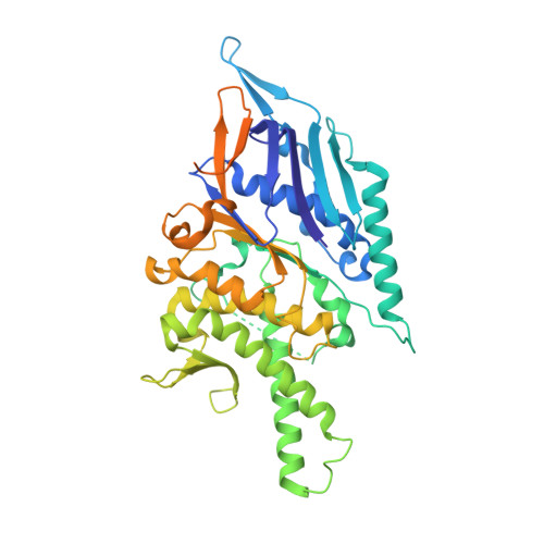
Escherichia coli Septu system, an anti-phage defense system, comprises two components: PtuA and PtuB. PtuA contains an ATPase domain, while PtuB is predicted to function as a nuclease. Here we show that PtuA and PtuB form a stable complex with a 6:2 stoichiometry. Cryo-electron microscopy structure of PtuAB reveals a distinctive horseshoe-like configuration. PtuA adopts a hexameric arrangement, organized as an asymmetric trimer of dimers, contrasting the ring-like structure by other ATPases. Notably, the three pairs of PtuA dimers assume distinct conformations and fulfill unique roles in recruiting PtuB. Our functional assays have further illuminated the importance of the oligomeric assembly of PtuAB in anti-phage defense. Moreover, we have uncovered that ATP molecules can directly bind to PtuA and inhibit the activities of PtuAB. Together, the assembly and function of the Septu system shed light on understanding other ATPase-containing systems in bacterial immunity.
- Department of Biotherapy, Cancer Center and State Key Laboratory of Biotherapy, West China Hospital, Sichuan University, Chengdu, P. R. China.
Organizational Affiliation: