Structural and spectroscopic characterization of RufO indicates a new biological role in rufomycin biosynthesis.
Jordan, S., Li, B., Traore, E., Wu, Y., Usai, R., Liu, A., Xie, Z.R., Wang, Y.(2023) J Biol Chem 299: 105049-105049
- PubMed: 37451485 Search on PubMedSearch on PubMed Central
- DOI: https://doi.org/10.1016/j.jbc.2023.105049
- Primary Citation Related Structures:
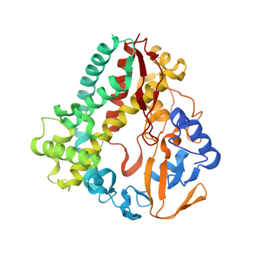
8SPP - PubMed Abstract:
Rufomycins constitute a class of cyclic heptapeptides isolated from actinomycetes. They are secondary metabolites that show promising treatment against Mycobacterium tuberculosis infections by inhibiting a novel drug target. Several nonproteinogenic amino acids are integrated into rufomycins, including a conserved 3-nitro-tyrosine. RufO, a cytochrome P450 (CYP)-like enzyme, was proposed to catalyze the formation of 3-nitro-tyrosine in the presence of O 2 and NO. To define its biological function, the interaction between RufO and the proposed substrate tyrosine is investigated using various spectroscopic methods that are sensitive to the structural change of a heme center. However, a low- to high-spin state transition and a dramatic increase in the redox potential that are commonly found in CYPs upon ligand binding have not been observed. Furthermore, a 1.89-Å crystal structure of RufO shows that the enzyme has flexible surface regions, a wide-open substrate access tunnel, and the heme center is largely exposed to solvent. Comparison with a closely related nitrating CYP reveals a spacious and hydrophobic distal pocket in RufO, which is incapable of stabilizing a free amino acid. Molecular docking validates the experimental data and proposes a possible substrate. Collectively, our results disfavor tyrosine as the substrate of RufO and point to the possibility that the nitration occurs during or after the assembly of the peptides. This study indicates a new function of the unique nitrating enzyme and provides insights into the biosynthesis of nonribosomal peptides.
- Department of Chemistry, University of Georgia, Athens, Georgia, USA.
Organizational Affiliation: