8SP7 | pdb_00008sp7

LINE-1 retrotransposon endonuclease domain complex with tranexamic acid


Experimental Data Snapshot

  • Method: X-RAY DIFFRACTION
  • Resolution: 1.83 Å
  • R-Value Free: 
    0.256 (Depositor), 0.256 (DCC) 
  • R-Value Work: 
    0.220 (Depositor), 0.220 (DCC) 
  • R-Value Observed: 
    0.222 (Depositor) 

Starting Model: experimental
View more details

wwPDB Validation 3D Report Full Report

Validation slider image for 8SP7

Ligand Structure Quality Assessment 


This is version 1.0 of the entry. See complete history

Literature

Identification and characterization of small molecule inhibitors of the LINE-1 retrotransposon endonuclease.

D'Ordine, A.M.Jogl, G.Sedivy, J.M.

(2024) Nat Commun 15: 3883-3883

  • DOI: https://doi.org/10.1038/s41467-024-48066-x
  • Primary Citation Related Structures: 
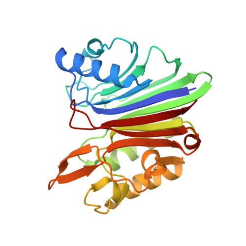
    8SP5, 8SP7

  • PubMed Abstract: 

    The long interspersed nuclear element-1 (LINE-1 or L1) retrotransposon is the only active autonomously replicating retrotransposon in the human genome. L1 harms the cell by inserting new copies, generating DNA damage, and triggering inflammation. Therefore, L1 inhibition could be used to treat many diseases associated with these processes. Previous research has focused on inhibition of the L1 reverse transcriptase due to the prevalence of well-characterized inhibitors of related viral enzymes. Here we present the L1 endonuclease as another target for reducing L1 activity. We characterize structurally diverse small molecule endonuclease inhibitors using computational, biochemical, and biophysical methods. We also show that these inhibitors reduce L1 retrotransposition, L1-induced DNA damage, and inflammation reinforced by L1 in senescent cells. These inhibitors could be used for further pharmacological development and as tools to better understand the life cycle of this element and its impact on disease processes.


  • Organizational Affiliation
    • Department of Molecular Biology, Cell Biology, and Biochemistry, Brown University, Providence, RI, USA.

Macromolecule Content 

  • Total Structure Weight: 27.5 kDa 
  • Atom Count: 1,896 
  • Modeled Residue Count: 234 
  • Deposited Residue Count: 239 
  • Unique protein chains: 1

Macromolecules

Find similar proteins by:|  3D Structure
Entity ID: 1
MoleculeChains  Sequence LengthOrganismDetailsImage
LINE-1 retrotransposon endonuclease239Homo sapiensMutation(s): 0 
EC: 2.7.7.49 (UniProt), 3.1.21 (UniProt)
UniProt
Find proteins for O00370 (Homo sapiens)
Explore O00370 
Go to UniProtKB:  O00370
Entity Groups
Sequence Clusters30% Identity50% Identity70% Identity90% Identity95% Identity100% Identity
UniProt GroupO00370
Sequence Annotations
Expand
Reference Sequence

Small Molecules

Experimental Data & Validation

Experimental Data

  • Method: X-RAY DIFFRACTION
  • Resolution: 1.83 Å
  • R-Value Free:  0.256 (Depositor), 0.256 (DCC) 
  • R-Value Work:  0.220 (Depositor), 0.220 (DCC) 
  • R-Value Observed: 0.222 (Depositor) 
Space Group: C 2 2 21
Unit Cell:
Length ( Å )Angle ( ˚ )
a = 54.222α = 90
b = 64.671β = 90
c = 132.358γ = 90
Software Package:
Software NamePurpose
PHENIXrefinement
XDSdata reduction
Aimlessdata scaling
PHASERphasing
EM Software:
TaskSoftware PackageVersion
MODEL REFINEMENTPHENIX1.21rc1_4895

Structure Validation

View Full Validation Report


Ligand Structure Quality Assessment 


Entry History 

& Funding Information

Deposition Data


Funding OrganizationLocationGrant Number
National Institutes of Health/National Institute on Aging (NIH/NIA)United States--

Revision History  (Full details and data files)

  • Version 1.0: 2024-05-22
    Type: Initial release