8SOZ

Structure of the complex formed by human interleukin-2 and scFv 602

  • Classification: CYTOKINE
  • Organism(s): Homo sapiens
  • Expression System: Homo sapiens
  • Mutation(s): No 

  • Deposited: 2023-04-30 Released: 2024-09-04 
  • Deposition Author(s): Gould, J.R., Leonard, E.K., Cao, S.D., Spangler, J.B.
  • Funding Organization(s): Department of Defense (DOD, United States), National Institutes of Health/National Cancer Institute (NIH/NCI), National Institutes of Health/National Institute of Biomedical Imaging and Bioengineering (NIH/NIBIB), V Foundation for Cancer Research, American Cancer Society, Maryland Stem Cell Research Fund, Emerson Collective, Willowcroft Foundation, Johns Hopkins University

Experimental Data Snapshot

  • Method: X-RAY DIFFRACTION
  • Resolution: 1.64 Å
  • R-Value Free: 0.250 
  • R-Value Work: 0.219 

Starting Models: experimental
View more details

wwPDB Validation   3D Report Full Report


This is version 1.2 of the entry. See complete history


Literature

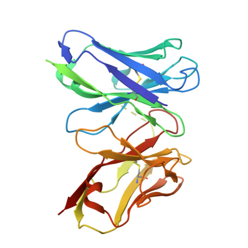
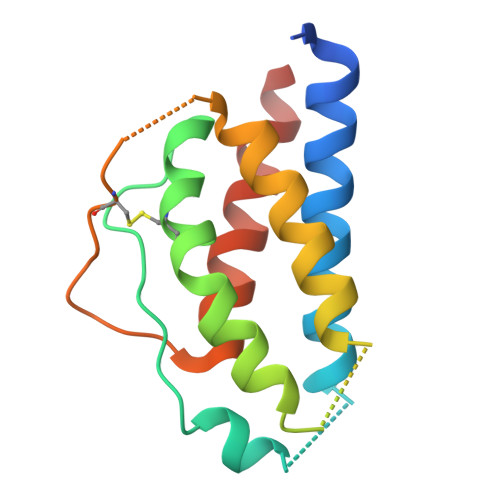
Engineered cytokine/antibody fusion proteins improve IL-2 delivery to pro-inflammatory cells and promote antitumor activity.

Leonard, E.K.Tomala, J.Gould, J.R.Leff, M.I.Lin, J.X.Li, P.Porter, M.J.Johansen, E.R.Thompson, L.Cao, S.D.Hou, S.Henclova, T.Huliciak, M.Sargunas, P.R.Fabilane, C.S.Vanek, O.Kovar, M.Schneider, B.Raimondi, G.Leonard, W.J.Spangler, J.B.

(2024) JCI Insight 9

  • DOI: https://doi.org/10.1172/jci.insight.173469
  • Primary Citation of Related Structures:  
    8SOW, 8SOZ

  • PubMed Abstract: 

    Progress in cytokine engineering is driving therapeutic translation by overcoming these proteins' limitations as drugs. The interleukin-2 (IL-2) cytokine is a promising immune stimulant for cancer treatment but is limited by its concurrent activation of both pro-inflammatory immune effector cells and anti-inflammatory regulatory T cells, toxicity at high doses, and short serum half-life. One approach to improve the selectivity, safety, and longevity of IL-2 is complexation with anti-IL-2 antibodies that bias the cytokine towards immune effector cell activation. Although this strategy shows potential in preclinical models, clinical translation of a cytokine/antibody complex is complicated by challenges in formulating a multi-protein drug and concerns regarding complex stability. Here, we introduced a versatile approach to designing intramolecularly assembled single-agent fusion proteins (immunocytokines, ICs) comprising IL-2 and a biasing anti-IL-2 antibody that directs the cytokine towards immune effector cells. We optimized IC construction and engineered the cytokine/antibody affinity to improve immune bias. We demonstrated that our IC preferentially activates and expands immune effector cells, leading to superior antitumor activity compared to natural IL-2, both alone and combined with immune checkpoint inhibitors. Moreover, therapeutic efficacy was observed without inducing toxicity. This work presents a roadmap for the design and translation of cytokine/antibody fusion proteins.


  • Organizational Affiliation

    Biomedical Engineering, Johns Hopkins University School of Medicine, Baltimore, United States of America.


Macromolecules
Find similar proteins by:  (by identity cutoff)  |  3D Structure
Entity ID: 1
MoleculeChains Sequence LengthOrganismDetailsImage
602 single chain fragment variableA [auth C]241Homo sapiensMutation(s): 0 
Entity Groups  
Sequence Clusters30% Identity50% Identity70% Identity90% Identity95% Identity100% Identity
Sequence Annotations
Expand
  • Reference Sequence
Find similar proteins by:  (by identity cutoff)  |  3D Structure
Entity ID: 2
MoleculeChains Sequence LengthOrganismDetailsImage
Interleukin-2B [auth A]139Homo sapiensMutation(s): 0 
Gene Names: IL2
UniProt & NIH Common Fund Data Resources
Find proteins for P60568 (Homo sapiens)
Explore P60568 
Go to UniProtKB:  P60568
PHAROS:  P60568
GTEx:  ENSG00000109471 
Entity Groups  
Sequence Clusters30% Identity50% Identity70% Identity90% Identity95% Identity100% Identity
UniProt GroupP60568
Sequence Annotations
Expand
  • Reference Sequence
Experimental Data & Validation

Experimental Data

  • Method: X-RAY DIFFRACTION
  • Resolution: 1.64 Å
  • R-Value Free: 0.250 
  • R-Value Work: 0.219 
  • Space Group: C 1 2 1
Unit Cell:
Length ( Å )Angle ( ˚ )
a = 137.192α = 90
b = 36.419β = 108.12
c = 64.189γ = 90
Software Package:
Software NamePurpose
PHENIXrefinement
XDSdata scaling
PHASERphasing
autoPROCdata reduction

Structure Validation

View Full Validation Report


Entry History & Funding Information

Deposition Data


Funding OrganizationLocationGrant Number
Department of Defense (DOD, United States)United StatesW81XWH1810735
Department of Defense (DOD, United States)United StatesW81XWH21P0031
National Institutes of Health/National Cancer Institute (NIH/NCI)United StatesR21CA249381
National Institutes of Health/National Institute of Biomedical Imaging and Bioengineering (NIH/NIBIB)United StatesR01EB029455
National Institutes of Health/National Institute of Biomedical Imaging and Bioengineering (NIH/NIBIB)United StatesR01EB029341
V Foundation for Cancer ResearchUnited StatesV2018-005
American Cancer SocietyUnited StatesPF-22-050-01-IBCD
Maryland Stem Cell Research FundUnited States2019-MSCRFD-5039
Emerson CollectiveUnited StatesCancer Research Fund
Willowcroft FoundationUnited States--
Johns Hopkins UniversityUnited StatesCatalyst award

Revision History  (Full details and data files)

  • Version 1.0: 2024-09-04
    Type: Initial release
  • Version 1.1: 2024-10-02
    Changes: Database references
  • Version 1.2: 2024-10-16
    Changes: Structure summary