Structure, dynamics, and redox reactivity of an all-purpose flavodoxin.
Khan, S., Ansari, A., Brachi, M., Das, D., El Housseini, W., Minteer, S., Miller, A.F.(2024) J Biol Chem 300: 107122-107122
- PubMed: 38417793 Search on PubMedSearch on PubMed Central
- DOI: https://doi.org/10.1016/j.jbc.2024.107122
- Primary Citation Related Structures:
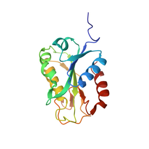
8SNZ, 8V2Y - PubMed Abstract:
The flavodoxin of Rhodopseudomonas palustris CGA009 (Rp9Fld) supplies highly reducing equivalents to crucial enzymes such as hydrogenase, especially when the organism is iron-restricted. By acquiring those electrons from photodriven electron flow via the bifurcating electron transfer flavoprotein, Rp9Fld provides solar power to vital metabolic processes. To understand Rp9Fld's ability to work with diverse partners, we solved its crystal structure. We observed the canonical flavodoxin (Fld) fold and features common to other long-chain Flds but not all the surface loops thought to recognize partner proteins. Moreover, some of the loops display alternative structures and dynamics. To advance studies of protein-protein associations and conformational consequences, we assigned the 19 F NMR signals of all five tyrosines (Tyrs). Our electrochemical measurements show that incorporation of 3- 19 F-Tyr in place of Tyr has only a modest effect on Rp9Fld's redox properties even though Tyrs flank the flavin on both sides. Meanwhile, the 19 F probes demonstrate the expected paramagnetic effect, with signals from nearby Tyrs becoming broadened beyond detection when the flavin semiquinone is formed. However, the temperature dependencies of chemical shifts and linewidths reveal dynamics affecting loops close to the flavin and regions that bind to partners in a variety of systems. These coincide with patterns of amino acid type conservation but not retention of specific residues, arguing against detailed specificity with respect to partners. We propose that the loops surrounding the flavin adopt altered conformations upon binding to partners and may even participate actively in electron transfer.
- Department of Chemistry, University of Kentucky, Lexington, Kentucky, USA.
Organizational Affiliation: