8SKW

MicroED structure of d(CGCGCG)2 Z-DNA


Experimental Data Snapshot

  • Method: ELECTRON CRYSTALLOGRAPHY
  • Resolution: 1.10 Å
  • R-Value Free: 0.226 
  • R-Value Work: 0.210 
  • R-Value Observed: 0.210 

wwPDB Validation   3D Report Full Report


This is version 1.1 of the entry. See complete history


Literature

Structure determination of a DNA crystal by MicroED.

Haymaker, A.Bardin, A.A.Gonen, T.Martynowycz, M.W.Nannenga, B.L.

(2023) Structure 31: 1499-1503.e2

  • DOI: https://doi.org/10.1016/j.str.2023.07.005
  • Primary Citation of Related Structures:  
    8SKW

  • PubMed Abstract: 

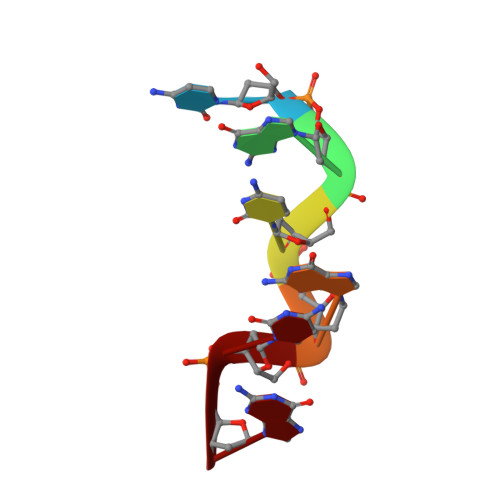
    Microcrystal electron diffraction (MicroED) is a powerful tool for determining high-resolution structures of microcrystals from a diverse array of biomolecular, chemical, and material samples. In this study, we apply MicroED to DNA crystals, which have not been previously analyzed using this technique. We utilized the d(CGCGCG) 2 DNA duplex as a model sample and employed cryo-FIB milling to create thin lamella for diffraction data collection. The MicroED data collection and subsequent processing resulted in a 1.10 Å resolution structure of the d(CGCGCG) 2 DNA, demonstrating the successful application of cryo-FIB milling and MicroED to the investigation of nucleic acid crystals.


  • Organizational Affiliation

    Biodesign Center for Applied Structural Discovery, Biodesign Institute, Arizona State University, 727 East Tyler Street, Tempe, AZ 85287, USA; School for Engineering of Matter, Transport and Energy, Arizona State University, Tempe, AZ, USA.


Macromolecules

Find similar nucleic acids by:  Sequence   |   3D Structure  

Entity ID: 1
MoleculeChains LengthOrganismImage
DNA (5'-D(*CP*GP*CP*GP*CP*G)-3')A [auth B],
B [auth A]
6synthetic construct
Sequence Annotations
Expand
  • Reference Sequence
Small Molecules
Ligands 1 Unique
IDChains Name / Formula / InChI Key2D Diagram3D Interactions
SPM
Query on SPM

Download Ideal Coordinates CCD File 
C [auth A]SPERMINE
C10 H26 N4
PFNFFQXMRSDOHW-UHFFFAOYSA-N
Experimental Data & Validation

Experimental Data

  • Method: ELECTRON CRYSTALLOGRAPHY
  • Resolution: 1.10 Å
  • R-Value Free: 0.226 
  • R-Value Work: 0.210 
  • R-Value Observed: 0.210 
  • Space Group: P 21 21 21
Unit Cell:
Length ( Å )Angle ( ˚ )
a = 18.28α = 90
b = 32β = 90
c = 41.67γ = 90
Software Package:
Software NamePurpose
phenix.refinerefinement
PHENIXrefinement

Structure Validation

View Full Validation Report


Entry History & Funding Information

Deposition Data


Funding OrganizationLocationGrant Number
National Institutes of Health/National Institute of General Medical Sciences (NIH/NIGMS)United StatesGM136508

Revision History  (Full details and data files)

  • Version 1.0: 2023-08-16
    Type: Initial release
  • Version 1.1: 2023-12-20
    Changes: Database references