8SKG | pdb_00008skg

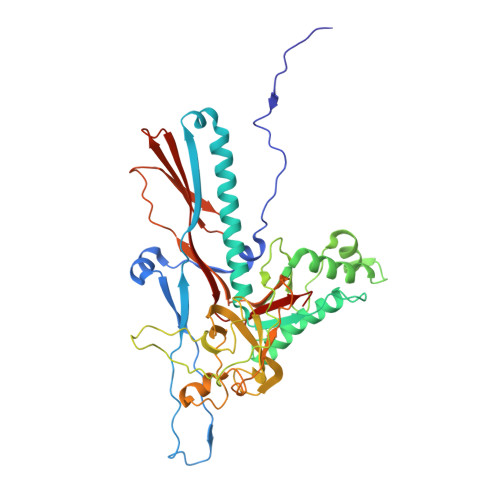
CUS-3 Mature Virus Capsid


Experimental Data Snapshot

  • Method: ELECTRON MICROSCOPY
  • Resolution: 2.80 Å
  • Aggregation State: PARTICLE 
  • Reconstruction Method: SINGLE PARTICLE 

wwPDB Validation 3D Report Full Report

Validation slider image for 8SKG

This is version 1.0 of the entry. See complete history

Literature

Not currently available

Whitehead III, R.D.White, S.Alexandrescu, A.T.Teschke, C.M.

To be published.

Macromolecule Content 

  • Total Structure Weight: 313.41 kDa 
  • Atom Count: 21,987 
  • Modeled Residue Count: 2,912 
  • Deposited Residue Count: 2,912 
  • Unique protein chains: 1

Macromolecules

Find similar proteins by:|  3D Structure
Entity ID: 1
MoleculeChains  Sequence LengthOrganismDetailsImage
Putative coat protein416Enterobacteria phage CUS-3Mutation(s): 0 
UniProt
Find proteins for A0ACD6B8E4 (Enterobacteria phage CUS-3)
Explore A0ACD6B8E4 
Go to UniProtKB:  A0ACD6B8E4
Entity Groups
Sequence Clusters30% Identity50% Identity70% Identity90% Identity95% Identity100% Identity
UniProt GroupA0ACD6B8E4
Sequence Annotations
Expand
Reference Sequence

Experimental Data & Validation

Experimental Data

  • Method: ELECTRON MICROSCOPY
  • Resolution: 2.80 Å
  • Aggregation State: PARTICLE 
  • Reconstruction Method: SINGLE PARTICLE 

Structure Validation

View Full Validation Report


Entry History 

& Funding Information

Deposition Data


Funding OrganizationLocationGrant Number
National Institutes of Health/National Institute of General Medical Sciences (NIH/NIGMS)United StatesR01 GM076661

Revision History  (Full details and data files)

  • Version 1.0: 2025-02-19
    Type: Initial release