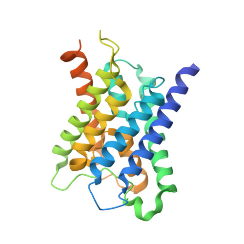
8SJY

Structure of lens aquaporin-0 array in sphingomyelin/cholesterol bilayer (1SM:2Chol)

  • Classification: MEMBRANE PROTEIN
  • Organism(s): Ovis aries
  • Mutation(s): No 
  • Membrane Protein: Yes  PDBTM

  • Deposited: 2023-04-18 Released: 2024-04-24 
  • Deposition Author(s): Chiu, P.-L., Walz, T.
  • Funding Organization(s): National Institutes of Health/National Eye Institute (NIH/NEI)

Experimental Data Snapshot

  • Method: ELECTRON CRYSTALLOGRAPHY
  • Resolution: 2.35 Å
  • R-Value Free: 0.287 
  • R-Value Work: 0.262 
  • R-Value Observed: 0.264 

Starting Model: experimental
View more details

wwPDB Validation   3D Report Full Report


This is version 1.1 of the entry. See complete history


Literature

Structure and dynamics of cholesterol-mediated aquaporin-0 arrays and implications for lipid rafts.

Chiu, P.L.Orjuela, J.D.de Groot, B.L.Aponte Santamaria, C.Walz, T.

(2024) Elife 12

  • DOI: https://doi.org/10.7554/eLife.90851
  • Primary Citation of Related Structures:  
    8SJX, 8SJY

  • PubMed Abstract: 

    Aquaporin-0 (AQP0) tetramers form square arrays in lens membranes through a yet unknown mechanism, but lens membranes are enriched in sphingomyelin and cholesterol. Here, we determined electron crystallographic structures of AQP0 in sphingomyelin/cholesterol membranes and performed molecular dynamics (MD) simulations to establish that the observed cholesterol positions represent those seen around an isolated AQP0 tetramer and that the AQP0 tetramer largely defines the location and orientation of most of its associated cholesterol molecules. At a high concentration, cholesterol increases the hydrophobic thickness of the annular lipid shell around AQP0 tetramers, which may thus cluster to mitigate the resulting hydrophobic mismatch. Moreover, neighboring AQP0 tetramers sandwich a cholesterol deep in the center of the membrane. MD simulations show that the association of two AQP0 tetramers is necessary to maintain the deep cholesterol in its position and that the deep cholesterol increases the force required to laterally detach two AQP0 tetramers, not only due to protein-protein contacts but also due to increased lipid-protein complementarity. Since each tetramer interacts with four such 'glue' cholesterols, avidity effects may stabilize larger arrays. The principles proposed to drive AQP0 array formation could also underlie protein clustering in lipid rafts.


  • Organizational Affiliation

    Department of Cell Biology, Harvard Medical School, Boston, United States.


Macromolecules
Find similar proteins by:  (by identity cutoff)  |  3D Structure
Entity ID: 1
MoleculeChains Sequence LengthOrganismDetailsImage
Lens fiber major intrinsic protein263Ovis ariesMutation(s): 0 
Membrane Entity: Yes 
UniProt
Find proteins for Q6J8I9 (Ovis aries)
Explore Q6J8I9 
Go to UniProtKB:  Q6J8I9
Entity Groups  
Sequence Clusters30% Identity50% Identity70% Identity90% Identity95% Identity100% Identity
UniProt GroupQ6J8I9
Sequence Annotations
Expand
  • Reference Sequence
Small Molecules
Ligands 2 Unique
IDChains Name / Formula / InChI Key2D Diagram3D Interactions
HWP (Subject of Investigation/LOI)
Query on HWP

Download Ideal Coordinates CCD File 
B [auth A],
C [auth A],
D [auth A],
E [auth A],
F [auth A]
[(E,2S,3R)-2-(hexadecanoylamino)-3-oxidanyl-octadec-4-enyl] 2-(trimethylazaniumyl)ethyl phosphate
C39 H79 N2 O6 P
RWKUXQNLWDTSLO-GWQJGLRPSA-N
CLR (Subject of Investigation/LOI)
Query on CLR

Download Ideal Coordinates CCD File 
G [auth A],
H [auth A],
I [auth A],
J [auth A]
CHOLESTEROL
C27 H46 O
HVYWMOMLDIMFJA-DPAQBDIFSA-N
Experimental Data & Validation

Experimental Data

  • Method: ELECTRON CRYSTALLOGRAPHY
  • Resolution: 2.35 Å
  • R-Value Free: 0.287 
  • R-Value Work: 0.262 
  • R-Value Observed: 0.264 
  • Space Group: P 4 2 2
Unit Cell:
Length ( Å )Angle ( ˚ )
a = 65.5α = 90
b = 65.5β = 90
c = 200γ = 90
Software Package:
Software NamePurpose
phenix.refinerefinement
PHENIXrefinement
PHASERphasing
IPLTdata reduction
EM Software:
TaskSoftware PackageVersion
RECONSTRUCTIONIPET0.9.8
MODEL REFINEMENTCNS1.3
MODEL REFINEMENTPHENIX1.20.1

Structure Validation

View Full Validation Report


Entry History & Funding Information

Deposition Data


Funding OrganizationLocationGrant Number
National Institutes of Health/National Eye Institute (NIH/NEI)United StatesEY015107

Revision History  (Full details and data files)

  • Version 1.0: 2024-04-24
    Type: Initial release
  • Version 1.1: 2024-09-25
    Changes: Data processing, Database references, Refinement description