


Crystal structure of HPPK from Methanocaldococcus jannaschii
Shaw, G.X., Needle, D., Stair, N.R., Cherry, S., Tropea, J.E., Waugh, D.S., Ji, X.To be published.
Experimental Data Snapshot
Starting Model: experimental
View more details
wwPDB Validation 3D Report Full Report

Entity ID: 1 | |||||
|---|---|---|---|---|---|
| Molecule | Chains | Sequence Length | Organism | Details | Image |
| 6-hydroxymethyl-7,8-dihydropterin pyrophosphokinase | 241 | Methanocaldococcus jannaschii | Mutation(s): 0 Gene Names: mptE, MJ1634 EC: 2.7.6.3 | 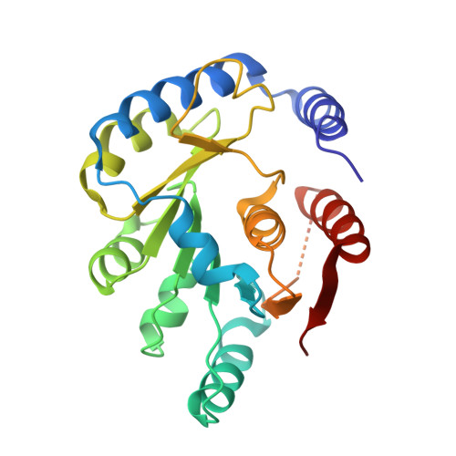 | |
UniProt | |||||
Entity Groups | |||||
| Sequence Clusters | 30% Identity50% Identity70% Identity90% Identity95% Identity100% Identity | ||||
| UniProt Group | Q59028 | ||||
Sequence AnnotationsExpand | |||||
Reference Sequence | |||||
| Ligands 1 Unique | |||||
|---|---|---|---|---|---|
| ID | Chains | Name / Formula / InChI Key | 2D Diagram | 3D Interactions | |
| SO4 Download:Ideal Coordinates CCD File | E [auth A], F [auth B], G [auth C] | SULFATE ION O4 S QAOWNCQODCNURD-UHFFFAOYSA-L |  | ||
| Length ( Å ) | Angle ( ˚ ) |
|---|---|
| a = 73.729 | α = 90 |
| b = 75.744 | β = 90 |
| c = 179.578 | γ = 90 |
| Software Name | Purpose |
|---|---|
| PHENIX | refinement |
| SCALEPACK | data scaling |
| DENZO | data reduction |
| PHASER | phasing |
| Funding Organization | Location | Grant Number |
|---|---|---|
| National Institutes of Health/National Cancer Institute (NIH/NCI) | United States | Intramural Research Program |


