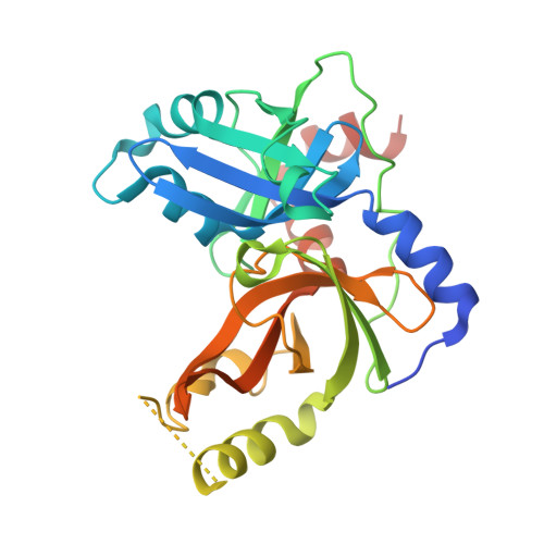
8S9K

Structure of dimeric FAM111A SPD S541A Mutant


Experimental Data Snapshot

  • Method: X-RAY DIFFRACTION
  • Resolution: 2.72 Å
  • R-Value Free: 0.249 
  • R-Value Work: 0.193 
  • R-Value Observed: 0.196 

Starting Model: in silico
View more details

wwPDB Validation   3D Report Full Report


This is version 1.0 of the entry. See complete history


Literature

Dimerization-dependent serine protease activity of FAM111A prevents replication fork stalling at topoisomerase 1 cleavage complexes.

Palani, S.Machida, Y.Alvey, J.R.Mishra, V.Welter, A.L.Cui, G.Bragantini, B.Botuyan, M.V.Cong, A.T.Q.Mer, G.Schellenberg, M.J.Machida, Y.J.

(2024) Nat Commun 15: 2064-2064

  • DOI: https://doi.org/10.1038/s41467-024-46207-w
  • Primary Citation of Related Structures:  
    8S9K, 8S9L

  • PubMed Abstract: 

    FAM111A, a serine protease, plays roles in DNA replication and antiviral defense. Missense mutations in the catalytic domain cause hyper-autocleavage and are associated with genetic disorders with developmental defects. Despite the enzyme's biological significance, the molecular architecture of the FAM111A serine protease domain (SPD) is unknown. Here, we show that FAM111A is a dimerization-dependent protease containing a narrow, recessed active site that cleaves substrates with a chymotrypsin-like specificity. X-ray crystal structures and mutagenesis studies reveal that FAM111A dimerizes via the N-terminal helix within the SPD. This dimerization induces an activation cascade from the dimerization sensor loop to the oxyanion hole through disorder-to-order transitions. Dimerization is essential for proteolytic activity in vitro and for facilitating DNA replication at DNA-protein crosslink obstacles in cells, while it is dispensable for autocleavage. These findings underscore the role of dimerization in FAM111A's function and highlight the distinction in its dimerization dependency between substrate cleavage and autocleavage.


  • Organizational Affiliation

    Mayo Clinic Graduate School of Biomedical Sciences, Mayo Clinic, Rochester, MN, USA.


Macromolecules
Find similar proteins by:  (by identity cutoff)  |  3D Structure
Entity ID: 1
MoleculeChains Sequence LengthOrganismDetailsImage
Serine protease FAM111A
A, B, C, D
280Homo sapiensMutation(s): 1 
Gene Names: FAM111AKIAA1895
EC: 3.4.21
UniProt & NIH Common Fund Data Resources
Find proteins for Q96PZ2 (Homo sapiens)
Explore Q96PZ2 
Go to UniProtKB:  Q96PZ2
PHAROS:  Q96PZ2
GTEx:  ENSG00000166801 
Entity Groups  
Sequence Clusters30% Identity50% Identity70% Identity90% Identity95% Identity100% Identity
UniProt GroupQ96PZ2
Sequence Annotations
Expand
  • Reference Sequence
Small Molecules
Ligands 1 Unique
IDChains Name / Formula / InChI Key2D Diagram3D Interactions
GOL
Query on GOL

Download Ideal Coordinates CCD File 
E [auth B]GLYCEROL
C3 H8 O3
PEDCQBHIVMGVHV-UHFFFAOYSA-N
Experimental Data & Validation

Experimental Data

  • Method: X-RAY DIFFRACTION
  • Resolution: 2.72 Å
  • R-Value Free: 0.249 
  • R-Value Work: 0.193 
  • R-Value Observed: 0.196 
  • Space Group: P 21 21 21
Unit Cell:
Length ( Å )Angle ( ˚ )
a = 74.814α = 90
b = 83.765β = 90
c = 191.12γ = 90
Software Package:
Software NamePurpose
PHENIXrefinement
SCALEPACKdata scaling
DENZOdata reduction
PHASERphasing

Structure Validation

View Full Validation Report


Entry History & Funding Information

Deposition Data


Funding OrganizationLocationGrant Number
National Institutes of Health/National Cancer Institute (NIH/NCI)United StatesCA233700
National Institutes of Health/National Cancer Institute (NIH/NCI)United StatesZIA BC 012086

Revision History  (Full details and data files)

  • Version 1.0: 2024-03-20
    Type: Initial release