8S7G | pdb_00008s7g

Cryo-EM structure of Pseudomonas aeruginosa Recombinase A (RecA) in complex with LexAS125A mutant


Experimental Data Snapshot

  • Method: ELECTRON MICROSCOPY
  • Resolution: 3.43 Å
  • Aggregation State: HELICAL ARRAY 
  • Reconstruction Method: SINGLE PARTICLE 

wwPDB Validation 3D Report Full Report

Validation slider image for 8S7G

This is version 1.3 of the entry. See complete history

Literature

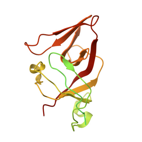
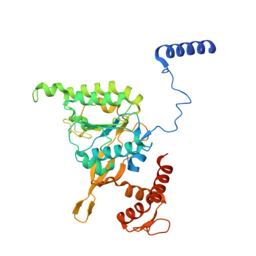
Snapshots of Pseudomonas aeruginosa SOS response reveal structural requisites for LexA autoproteolysis.

Vascon, F.De Felice, S.Gasparotto, M.Huber, S.T.Catalano, C.Chinellato, M.Mezzetti, R.Grinzato, A.Filippini, F.Maso, L.Jakobi, A.J.Cendron, L.

(2025) iScience 28: 111726-111726

  • DOI: https://doi.org/10.1016/j.isci.2024.111726
  • Primary Citation Related Structures: 
    8B0V, 8S70, 8S7G

  • PubMed Abstract: 

    Antimicrobial resistance poses a severe threat to human health and Pseudomonas aeruginosa stands out among the pathogens responsible for this emergency. The SOS response to DNA damage is crucial in bacterial evolution, influencing resistance development and adaptability in challenging environments, especially under antibiotic exposure. Recombinase A (RecA) and the transcriptional repressor LexA are the key players that orchestrate this process, determining either the silencing or the active transcription of the genes under their control. By integrating state-of-the-art structural approaches with in vitro binding and functional assays, we elucidated the molecular events activating the SOS response in P. aeruginosa , focusing on the RecA-LexA interaction. Our findings identify the conserved determinants and strength of the interactions that allow RecA to trigger LexA autocleavage and inactivation. These results provide the groundwork for designing novel antimicrobial strategies and exploring the potential translation of Escherichia coli -derived approaches, to address the implications of P. aeruginosa infections.


  • Organizational Affiliation
    • Department of Biology, University of Padua, Via Ugo Bassi 58/b, 35131 Padova, Italy.

Macromolecule Content 

  • Total Structure Weight: 491.04 kDa 
  • Atom Count: 30,036 
  • Modeled Residue Count: 3,892 
  • Deposited Residue Count: 4,429 
  • Unique protein chains: 2
  • Unique nucleic acid chains: 1

Macromolecules


Find similar proteins by:|  3D Structure
Entity ID: 1
MoleculeChains  Sequence LengthOrganismDetailsImage
LexA repressor
A, B
211Pseudomonas aeruginosaMutation(s): 1 
Gene Names: lexAPA3007
EC: 3.4.21.88
UniProt
Find proteins for P37452 (Pseudomonas aeruginosa (strain ATCC 15692 / DSM 22644 / CIP 104116 / JCM 14847 / LMG 12228 / 1C / PRS 101 / PAO1))
Explore P37452 
Go to UniProtKB:  P37452
Entity Groups
Sequence Clusters30% Identity50% Identity70% Identity90% Identity95% Identity100% Identity
UniProt GroupP37452
Sequence Annotations
Expand
Reference Sequence
Find similar proteins by:|  3D Structure
Entity ID: 2
MoleculeChains  Sequence LengthOrganismDetailsImage
Protein RecA361Pseudomonas aeruginosaMutation(s): 0 
Gene Names: recAPA3617
UniProt
Find proteins for P08280 (Pseudomonas aeruginosa (strain ATCC 15692 / DSM 22644 / CIP 104116 / JCM 14847 / LMG 12228 / 1C / PRS 101 / PAO1))
Explore P08280 
Go to UniProtKB:  P08280
Entity Groups
Sequence Clusters30% Identity50% Identity70% Identity90% Identity95% Identity100% Identity
UniProt GroupP08280
Sequence Annotations
Expand
Reference Sequence
Find similar nucleic acids by:  (by identity cutoff) 
Entity ID: 3
MoleculeChains LengthOrganismImage
DNA (36-MER)L [auth T]36synthetic construct
Sequence Annotations
Expand
Reference Sequence

Small Molecules

Ligands 2 Unique
IDChains Name / Formula / InChI Key2D Diagram3D Interactions
AGS
(Subject of Investigation/LOI)

Query on AGS


Download:Ideal Coordinates CCD File
BA [auth J]
DA [auth K]
FA [auth L]
HA [auth C]
JA [auth M]
BA [auth J],
DA [auth K],
FA [auth L],
HA [auth C],
JA [auth M],
P [auth D],
R [auth E],
T [auth F],
V [auth G],
X [auth H],
Z [auth I]
PHOSPHOTHIOPHOSPHORIC ACID-ADENYLATE ESTER
C10 H16 N5 O12 P3 S
NLTUCYMLOPLUHL-KQYNXXCUSA-N
MG
(Subject of Investigation/LOI)

Query on MG


Download:Ideal Coordinates CCD File
AA [auth J]
CA [auth K]
EA [auth L]
GA [auth C]
IA [auth M]
AA [auth J],
CA [auth K],
EA [auth L],
GA [auth C],
IA [auth M],
O [auth D],
Q [auth E],
S [auth F],
U [auth G],
W [auth H],
Y [auth I]
MAGNESIUM ION
Mg
JLVVSXFLKOJNIY-UHFFFAOYSA-N

Experimental Data & Validation

Experimental Data

  • Method: ELECTRON MICROSCOPY
  • Resolution: 3.43 Å
  • Aggregation State: HELICAL ARRAY 
  • Reconstruction Method: SINGLE PARTICLE 
EM Software:
TaskSoftware PackageVersion
RECONSTRUCTIONcryoSPARC4.2.1
MODEL REFINEMENTPHENIX

Structure Validation

View Full Validation Report


Entry History 

& Funding Information

Deposition Data


Funding OrganizationLocationGrant Number
Italian Ministry of EducationItaly--

Revision History  (Full details and data files)

  • Version 1.0: 2025-01-15
    Type: Initial release
  • Version 1.1: 2025-02-12
    Changes: Data collection, Database references
  • Version 1.2: 2025-03-12
    Changes: Data collection
  • Version 1.3: 2025-07-02
    Changes: Data collection