8S4T

DNA bound structure of PrgE from plasmid pCF10


Experimental Data Snapshot

  • Method: X-RAY DIFFRACTION
  • Resolution: 2.67 Å
  • R-Value Free: 0.253 
  • R-Value Work: 0.229 
  • R-Value Observed: 0.231 

Starting Model: in silico
View more details

wwPDB Validation   3D Report Full Report


This is version 1.1 of the entry. See complete history


Literature

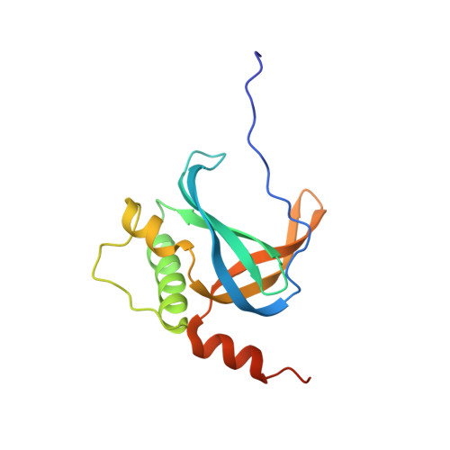
PrgE: an OB-fold protein from plasmid pCF10 with striking differences to prototypical bacterial SSBs.

Breidenstein, A.Lamy, A.Bader, C.P.Sun, W.S.Wanrooij, P.H.Berntsson, R.P.

(2024) Life Sci Alliance 7

  • DOI: https://doi.org/10.26508/lsa.202402693
  • Primary Citation of Related Structures:  
    8S4S, 8S4T

  • PubMed Abstract: 

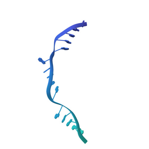
    A major pathway for horizontal gene transfer is the transmission of DNA from donor to recipient cells via plasmid-encoded type IV secretion systems (T4SSs). Many conjugative plasmids encode for a single-stranded DNA-binding protein (SSB) together with their T4SS. Some of these SSBs have been suggested to aid in establishing the plasmid in the recipient cell, but for many, their function remains unclear. Here, we characterize PrgE, a proposed SSB from the Enterococcus faecalis plasmid pCF10. We show that PrgE is not essential for conjugation. Structurally, it has the characteristic OB-fold of SSBs, but it has very unusual DNA-binding properties. Our DNA-bound structure shows that PrgE binds ssDNA like beads on a string supported by its N-terminal tail. In vitro studies highlight the plasticity of PrgE oligomerization and confirm the importance of the N-terminus. Unlike other SSBs, PrgE binds both double- and single-stranded DNA equally well. This shows that PrgE has a quaternary assembly and DNA-binding properties that are very different from the prototypical bacterial SSB, but also different from eukaryotic SSBs.


  • Organizational Affiliation

    https://ror.org/05kb8h459 Department of Medical Biochemistry and Biophysics, Umeå University, Umeå, Sweden.


Macromolecules

Find similar proteins by:  (by identity cutoff)  |  3D Structure
Entity ID: 1
MoleculeChains Sequence LengthOrganismDetailsImage
PrgE
A, B, C
145Enterococcus faecalisMutation(s): 0 
Gene Names: 
UniProt
Find proteins for D1LHE9 (Enterococcus faecalis)
Explore D1LHE9 
Go to UniProtKB:  D1LHE9
Entity Groups  
Sequence Clusters30% Identity50% Identity70% Identity90% Identity95% Identity100% Identity
UniProt GroupD1LHE9
Sequence Annotations
Expand
  • Reference Sequence
Find similar nucleic acids by:  (by identity cutoff)  |  3D Structure
Entity ID: 2
MoleculeChains LengthOrganismImage
DNA60DNA molecule
Sequence Annotations
Expand
  • Reference Sequence
Small Molecules
Experimental Data & Validation

Experimental Data

  • Method: X-RAY DIFFRACTION
  • Resolution: 2.67 Å
  • R-Value Free: 0.253 
  • R-Value Work: 0.229 
  • R-Value Observed: 0.231 
  • Space Group: P 21 21 21
Unit Cell:
Length ( Å )Angle ( ˚ )
a = 75.776α = 90
b = 90.043β = 90
c = 131.848γ = 90
Software Package:
Software NamePurpose
REFMACrefinement
XDSdata reduction
XDSdata scaling
PHENIXphasing
Cootmodel building

Structure Validation

View Full Validation Report


Entry History & Funding Information

Deposition Data


Funding OrganizationLocationGrant Number
Swedish Research CouncilSweden2016-03599
Knut and Alice Wallenberg FoundationSweden--
Carl Trygger FoundationSwedenCTS 18:39

Revision History  (Full details and data files)

  • Version 1.0: 2024-05-29
    Type: Initial release
  • Version 1.1: 2024-07-03
    Changes: Database references