Tetrahydropyridine LIMK inhibitors: Structure activity studies and biological characterization.
Champire, A., Berabez, R., Braka, A., Cosson, A., Corret, J., Girardin, C., Serrano, A., Aci-Seche, S., Bonnet, P., Josselin, B., Brindeau, P., Ruchaud, S., Leguevel, R., Chatterjee, D., Mathea, S., Knapp, S., Brion, R., Verrecchia, F., Vallee, B., Ple, K., Benedetti, H., Routier, S.(2024) Eur J Med Chem 271: 116391-116391
- PubMed: 38669909 Search on PubMed
- DOI: https://doi.org/10.1016/j.ejmech.2024.116391
- Primary Citation Related Structures:
8S3X - PubMed Abstract:
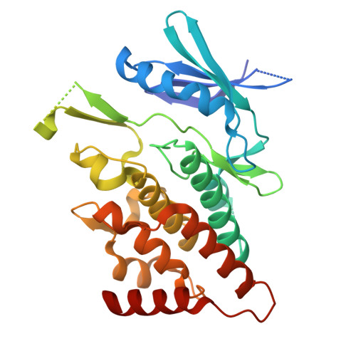
LIM Kinases, LIMK1 and LIMK2, have become promising targets for the development of inhibitors with potential application for the treatment of several major diseases. LIMKs play crucial roles in cytoskeleton remodeling as downstream effectors of small G proteins of the Rho-GTPase family, and as major regulators of cofilin, an actin depolymerizing factor. In this article we describe the conception, synthesis, and biological evaluation of novel tetrahydropyridine pyrrolopyrimidine LIMK inhibitors. Homology models were first constructed to better understand the binding mode of our preliminary compounds and to explain differences in biological activity. A library of over 60 products was generated and in vitro enzymatic activities were measured in the mid to low nanomolar range. The most promising derivatives were then evaluated in cell on cofilin phosphorylation inhibition which led to the identification of 52 which showed excellent selectivity for LIMKs in a kinase selectivity panel. We also demonstrated that 52 affected the cell cytoskeleton by disturbing actin filaments. Cell migration studies with this derivative using three different cell lines displayed a significant effect on cell motility. Finally, the crystal structure of the kinase domain of LIMK2 complexed with 52 was solved, greatly improving our understanding of the interaction between 52 and LIMK2 active site. The reported data represent a basis for the development of more efficient LIMK inhibitors for future in vivo preclinical validation.
- ICOA, Université d'Orléans, CNRS UMR 7311, 45067, Orléans, France.
Organizational Affiliation: