The oligomeric states of dye-decolorizing peroxidases from Streptomyces lividans and their implications for mechanism of substrate oxidation.
Lucic, M., Allport, T., Clarke, T.A., Williams, L.J., Wilson, M.T., Chaplin, A.K., Worrall, J.A.R.(2024) Protein Sci 33: e5073-e5073
- PubMed: 38864770 Search on PubMedSearch on PubMed Central
- DOI: https://doi.org/10.1002/pro.5073
- Primary Citation Related Structures:
8RWY - PubMed Abstract:
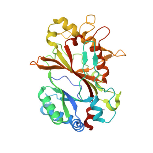
A common evolutionary mechanism in biology to drive function is protein oligomerization. In prokaryotes, the symmetrical assembly of repeating protein units to form homomers is widespread, yet consideration in vitro of whether such assemblies have functional or mechanistic consequences is often overlooked. Dye-decolorizing peroxidases (DyPs) are one such example, where their dimeric α + β barrel units can form various oligomeric states, but the oligomer influence, if any, on mechanism and function has received little attention. In this work, we have explored the oligomeric state of three DyPs found in Streptomyces lividans, each with very different mechanistic behaviors in their reactions with hydrogen peroxide and organic substrates. Using analytical ultracentrifugation, we reveal that except for one of the A-type DyPs where only a single sedimenting species is detected, oligomer states ranging from homodimers to dodecamers are prevalent in solution. Using cryo-EM on preparations of the B-type DyP, we determined a 3.02 Å resolution structure of a hexamer assembly that corresponds to the dominant oligomeric state in solution as determined by analytical ultracentrifugation. Furthermore, cryo-EM data detected sub-populations of higher-order oligomers, with one of these formed by an arrangement of two B-type DyP hexamers to give a dodecamer assembly. Our solution and structural insights of these oligomer states provide a new framework to consider previous mechanistic studies of these DyP members and are discussed in terms of long-range electron transfer for substrate oxidation and in the "storage" of oxidizable equivalents on the heme until a two-electron donor is available.
- School of Life Sciences, University of Essex, Colchester, UK.
Organizational Affiliation: