8RSN | pdb_00008rsn

macrodomain-fused SirTM (Mfs1) from Fusarium oxysporum f. sp. cubense race 1


Experimental Data Snapshot

  • Method: X-RAY DIFFRACTION
  • Resolution: 2.22 Å
  • R-Value Free: 
    0.253 (Depositor), 0.251 (DCC) 
  • R-Value Work: 
    0.203 (Depositor), 0.205 (DCC) 

Starting Model: in silico
View more details

wwPDB Validation 3D Report Full Report

Validation slider image for 8RSN

This is version 1.2 of the entry. See complete history

Literature

Evolutionary and molecular basis of ADP-ribosylation reversal by zinc-dependent macrodomains.

Ariza, A.Liu, Q.Cowieson, N.P.Ahel, I.Filippov, D.V.Rack, J.G.M.

(2024) J Biological Chem 300: 107770-107770

  • DOI: https://doi.org/10.1016/j.jbc.2024.107770
  • Primary Citation Related Structures: 
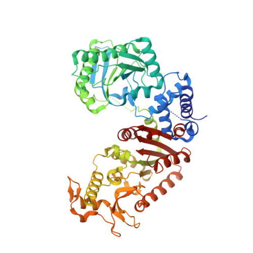
    8RSI, 8RSJ, 8RSK, 8RSL, 8RSM, 8RSN

  • PubMed Abstract: 

    Dynamic ADP-ribosylation signalling is a crucial pathway that controls fundamental cellular processes, in particular, the response to cellular stresses such as DNA damage, reactive oxygen species and infection. In some pathogenic microbes the response to oxidative stress is controlled by a SirTM/zinc-containing macrodomain (Zn-Macro) pair responsible for establishment and removal of the modification, respectively. Targeting this defence mechanism against the host's innate immune response may lead to novel approaches to support the fight against emerging antimicrobial resistance. Earlier studies suggested that Zn-Macros play a key role in the activation of this defence. Therefore, we used phylogenetic, biochemical, and structural approaches to elucidate the functional properties of these enzymes. Using the substrate mimetic asparagine-ADP-ribose as well as the ADP-ribose product, we characterise the catalytic role of the zinc ion in the removal of the ADP-ribosyl modification. Furthermore, we determined structural properties that contribute to substrate selectivity within the different Zn-Macro branches. Together, our data not only give new insights into the Zn-Macro family but also highlight their distinct features that may be exploited for the development of future therapies.


  • Organizational Affiliation
    • School of Biosciences, University of Sheffield, Alfred Denny Building, Western Bank, Sheffield, S10 2TN, UK; Sir William Dunn School of Pathology, University of Oxford, South Parks Road, Oxford, OX1 3RE, UK.

Macromolecule Content 

  • Total Structure Weight: 133.66 kDa 
  • Atom Count: 8,664 
  • Modeled Residue Count: 1,070 
  • Deposited Residue Count: 1,198 
  • Unique protein chains: 1

Macromolecules

Find similar proteins by:|  3D Structure
Entity ID: 1
MoleculeChains  Sequence LengthOrganismDetailsImage
ADP-ribose 1''-phosphate phosphatase
A, B
599Fusarium oxysporum f. sp. cubense race 1Mutation(s): 2 
Gene Names: Focb16_v015632
EC: 3.1.3.84 (PDB Primary Data), 2.4.2 (UniProt), 3.2.1 (UniProt)
UniProt
Find proteins for A0A559KX76 (Fusarium oxysporum f. sp. cubense)
Explore A0A559KX76 
Go to UniProtKB:  A0A559KX76
Entity Groups
Sequence Clusters30% Identity50% Identity70% Identity90% Identity95% Identity100% Identity
UniProt GroupA0A559KX76
Sequence Annotations
Expand
Reference Sequence

Small Molecules

Ligands 4 Unique
IDChains Name / Formula / InChI Key2D Diagram3D Interactions
ZN

Query on ZN


Download:Ideal Coordinates CCD File
L [auth A],
V [auth B]
ZINC ION
Zn
PTFCDOFLOPIGGS-UHFFFAOYSA-N
EDO

Query on EDO


Download:Ideal Coordinates CCD File
C [auth A]
D [auth A]
I [auth A]
J [auth A]
K [auth A]
C [auth A],
D [auth A],
I [auth A],
J [auth A],
K [auth A],
N [auth B],
O [auth B],
T [auth B]
1,2-ETHANEDIOL
C2 H6 O2
LYCAIKOWRPUZTN-UHFFFAOYSA-N
SCN

Query on SCN


Download:Ideal Coordinates CCD File
E [auth A]
F [auth A]
G [auth A]
H [auth A]
P [auth B]
E [auth A],
F [auth A],
G [auth A],
H [auth A],
P [auth B],
Q [auth B],
R [auth B],
S [auth B],
U [auth B]
THIOCYANATE ION
C N S
ZMZDMBWJUHKJPS-UHFFFAOYSA-M
K

Query on K


Download:Ideal Coordinates CCD File
M [auth A],
W [auth B]
POTASSIUM ION
K
NPYPAHLBTDXSSS-UHFFFAOYSA-N

Experimental Data & Validation

Experimental Data

  • Method: X-RAY DIFFRACTION
  • Resolution: 2.22 Å
  • R-Value Free:  0.253 (Depositor), 0.251 (DCC) 
  • R-Value Work:  0.203 (Depositor), 0.205 (DCC) 
Space Group: P 21 21 21
Unit Cell:
Length ( Å )Angle ( ˚ )
a = 61.739α = 90
b = 135.089β = 90
c = 147.625γ = 90
Software Package:
Software NamePurpose
REFMACrefinement
DIALSdata reduction
Aimlessdata scaling
PHASERphasing

Structure Validation

View Full Validation Report


Entry History 

& Funding Information

Deposition Data

  • Released Date: 2024-09-25 
  • Deposition Author(s): Ariza, A.

Funding OrganizationLocationGrant Number
Wellcome TrustUnited Kingdom210634
Biotechnology and Biological Sciences Research Council (BBSRC)United KingdomBB/R007195/1
Biotechnology and Biological Sciences Research Council (BBSRC)United KingdomBB/W016613/1
Medical Research Council (MRC, United Kingdom)United KingdomMR/X007472/1

Revision History  (Full details and data files)

  • Version 1.0: 2024-09-25
    Type: Initial release
  • Version 1.1: 2024-10-02
    Changes: Database references
  • Version 1.2: 2024-10-16
    Changes: Database references, Structure summary