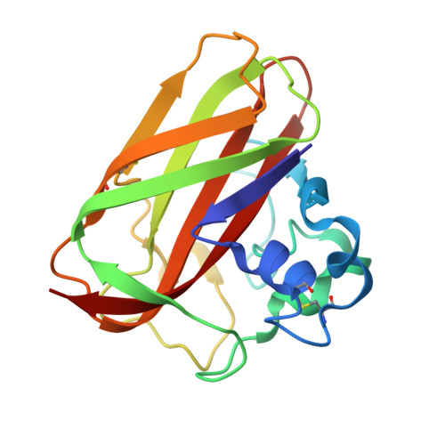
Integrated Experimental and Theoretical Investigation of Copper Active Site Properties of a Lytic Polysaccharide Monooxygenase from Serratia marcescens.
Munzone, A., Pujol, M., Tamhankar, A., Joseph, C., Mazurenko, I., Reglier, M., Jannuzzi, S.A.V., Royant, A., Sicoli, G., DeBeer, S., Orio, M., Simaan, A.J., Decroos, C.(2024) Inorg Chem 63: 11063-11078
- PubMed: 38814816
- DOI: https://doi.org/10.1021/acs.inorgchem.4c00602
- Primary Citation of Related Structures:
8RRY - PubMed Abstract:
In this paper, we employed a multidisciplinary approach, combining experimental techniques and density functional theory (DFT) calculations to elucidate key features of the copper coordination environment of the bacterial lytic polysaccharide monooxygenase (LPMO) from Serratia marcescens ( Sm AA10). The structure of the holo -enzyme was successfully obtained by X-ray crystallography. We then determined the copper(II) binding affinity using competing ligands and observed that the affinity of the histidine brace ligands for copper is significantly higher than previously described. UV-vis, advanced electron paramagnetic resonance (EPR), and X-ray absorption spectroscopy (XAS) techniques, including high-energy resolution fluorescence detected (HERFD) XAS, were further used to gain insight into the copper environment in both the Cu(II) and Cu(I) redox states. The experimental data were successfully rationalized by DFT models, offering valuable information on the electronic structure and coordination geometry of the copper center. Finally, the Cu(II)/Cu(I) redox potential was determined using two different methods at ca . 350 mV vs NHE and rationalized by DFT calculations. This integrated approach not only advances our knowledge of the active site properties of Sm AA10 but also establishes a robust framework for future studies of similar enzymatic systems.
Organizational Affiliation:
Aix Marseille Univ, CNRS, Centrale Méditerranée, iSm2, Marseille 13013, France.


