8RPX | pdb_00008rpx

NhoI restriction endonuclease in complex with quadruply methylated DNA target


Experimental Data Snapshot

  • Method: X-RAY DIFFRACTION
  • Resolution: 1.81 Å
  • R-Value Free: 
    0.243 (Depositor), 0.270 (DCC) 
  • R-Value Work: 
    0.205 (Depositor) 
  • R-Value Observed: 
    0.206 (Depositor) 

Starting Model: in silico
View more details

wwPDB Validation 3D Report Full Report

Validation slider image for 8RPX

This is version 1.2 of the entry. See complete history

Literature

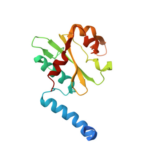
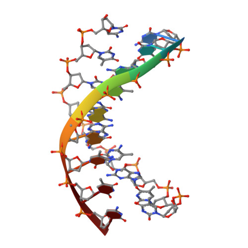
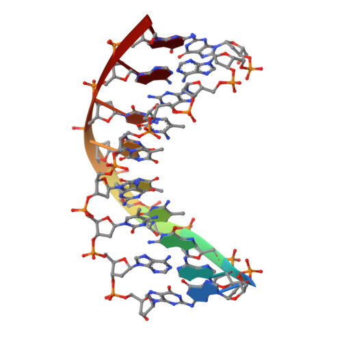
Structural analysis of the BisI family of modification dependent restriction endonucleases.

Szafran, K.Rafalski, D.Skowronek, K.Wojciechowski, M.Kazrani, A.A.Gilski, M.Xu, S.Y.Bochtler, M.

(2024) Nucleic Acids Res 52: 9103-9118

  • DOI: https://doi.org/10.1093/nar/gkae634
  • Primary Citation Related Structures: 
    8Q5M, 8Q5N, 8Q5O, 8RPX

  • PubMed Abstract: 

    The BisI family of restriction endonucleases is unique in requiring multiple methylated or hydroxymethylated cytosine residues within a short recognition sequence (GCNGC), and in cleaving directly within this sequence, rather than at a distance. Here, we report that the number of modified cytosines that are required for cleavage can be tuned by the salt concentration. We present crystal structures of two members of the BisI family, NhoI and Eco15I_Ntd (N-terminal domain of Eco15I), in the absence of DNA and in specific complexes with tetra-methylated GCNGC target DNA. The structures show that NhoI and Eco15I_Ntd sense modified cytosine bases in the context of double-stranded DNA (dsDNA) without base flipping. In the co-crystal structures of NhoI and Eco15I_Ntd with DNA, the internal methyl groups (G5mCNGC) interact with the side chains of an (H/R)(V/I/T/M) di-amino acid motif near the C-terminus of the distal enzyme subunit and arginine residue from the proximal subunit. The external methyl groups (GCNG5mC) interact with the proximal enzyme subunit, mostly through main chain contacts. Surface plasmon resonance analysis for Eco15I_Ntd shows that the internal and external methyl binding pockets contribute about equally to sensing of cytosine methyl groups.


  • Organizational Affiliation
    • International Institute of Molecular and Cell Biology, Warsaw, Poland.

Macromolecule Content 

  • Total Structure Weight: 113.9 kDa 
  • Atom Count: 8,612 
  • Modeled Residue Count: 851 
  • Deposited Residue Count: 910 
  • Unique protein chains: 1
  • Unique nucleic acid chains: 2

Macromolecules


Find similar proteins by:|  3D Structure
Entity ID: 1
MoleculeChains  Sequence LengthOrganismDetailsImage
Restriction endonuclease (NhoI)
A, B, C, D, E
171Nitrolancea hollandicaMutation(s): 0 
Gene Names: NITHO_2570002
UniProt
Find proteins for I4EG67 (Nitrolancea hollandica Lb)
Explore I4EG67 
Go to UniProtKB:  I4EG67
Entity Groups
Sequence Clusters30% Identity50% Identity70% Identity90% Identity95% Identity100% Identity
UniProt GroupI4EG67
Sequence Annotations
Expand
Reference Sequence
Find similar nucleic acids by:  Sequence
Entity ID: 2
MoleculeChains LengthOrganismImage
DNA (5'-D(*CP*TP*GP*(5CM)P*AP*GP*(5CM)P*TP*C)-3')
F, H, J
9synthetic construct
Sequence Annotations
Expand
Reference Sequence
Find similar nucleic acids by:  Sequence
Entity ID: 3
MoleculeChains LengthOrganismImage
DNA(5'-D(*GP*AP*GP*(5CM)P*TP*GP*(5CM)P*AP*GP)-3')
G, I, K
9synthetic construct
Sequence Annotations
Expand
Reference Sequence

Small Molecules

Ligands 2 Unique
IDChains Name / Formula / InChI Key2D Diagram3D Interactions
EDO

Query on EDO


Download:Ideal Coordinates CCD File
L [auth A]
N [auth B]
O [auth B]
R [auth D]
S [auth D]
L [auth A],
N [auth B],
O [auth B],
R [auth D],
S [auth D],
T [auth D],
U [auth D],
X [auth H]
1,2-ETHANEDIOL
C2 H6 O2
LYCAIKOWRPUZTN-UHFFFAOYSA-N
CA
(Subject of Investigation/LOI)

Query on CA


Download:Ideal Coordinates CCD File
M [auth A],
P [auth B],
Q [auth C],
V [auth D],
W [auth E]
CALCIUM ION
Ca
BHPQYMZQTOCNFJ-UHFFFAOYSA-N

Experimental Data & Validation

Experimental Data

  • Method: X-RAY DIFFRACTION
  • Resolution: 1.81 Å
  • R-Value Free:  0.243 (Depositor), 0.270 (DCC) 
  • R-Value Work:  0.205 (Depositor) 
  • R-Value Observed: 0.206 (Depositor) 
Space Group: P 21 21 2
Unit Cell:
Length ( Å )Angle ( ˚ )
a = 114.635α = 90
b = 167.059β = 90
c = 56.514γ = 90
Software Package:
Software NamePurpose
PHENIXrefinement
Cootmodel building
PHASERphasing
MOLREPphasing
XDSdata reduction
XDSdata scaling

Structure Validation

View Full Validation Report


Entry History 

& Funding Information

Deposition Data


Funding OrganizationLocationGrant Number
Other governmentPolandPPI/APM/2018/1/00034/U/001

Revision History  (Full details and data files)

  • Version 1.0: 2024-07-17
    Type: Initial release
  • Version 1.1: 2024-08-07
    Changes: Database references
  • Version 1.2: 2024-09-04
    Changes: Database references