8ROV | pdb_00008rov

Human dectin-2 with dimerization domain


Experimental Data Snapshot

  • Method: X-RAY DIFFRACTION
  • Resolution: 2.36 Å
  • R-Value Free: 
    0.284 (Depositor), 0.284 (DCC) 
  • R-Value Work: 
    0.222 (Depositor), 0.222 (DCC) 
  • R-Value Observed: 
    0.225 (Depositor) 

Starting Model: experimental
View more details

wwPDB Validation 3D Report Full Report

Validation slider image for 8ROV

Ligand Structure Quality Assessment 


This is version 1.1 of the entry. See complete history

Literature

Interactions that define the arrangement of sugar-binding sites in BDCA-2 and dectin-2 dimers.

Liu, Y.Kim, J.W.Feinberg, H.Cull, N.Weis, W.I.Taylor, M.E.Drickamer, K.

(2024) Glycobiology 34

  • DOI: https://doi.org/10.1093/glycob/cwae082
  • Primary Citation Related Structures: 
    8ROV

  • PubMed Abstract: 

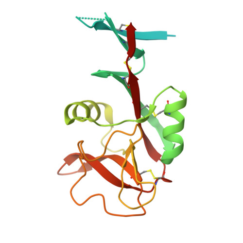
    The sugar-binding receptors dectin-2 and blood dendritic cell antigen 2 (BDCA-2) bind oligosaccharide ligands through extracellular carbohydrate-recognition domains (CRDs) and initiate intracellular signaling through Fc receptor γ adapters (FcRγ). Dectin-2 stimulates macrophages in response to pathogen binding while BDCA-2 modulates cytokine production in plasmacytoid dendritic cells. The oligomeric states of these receptors and the orientations of their CRDs have been investigated by analysis of a naturally occurring disulfide-bonded variant of BDCA-2 and by replacement of transmembrane domains with N-terminal dimerization domains to create extracellular domain dimers of both dectin-2 and BDCA-2. Analysis of these constructs, as well as previously described crystal structures of the CRDs from these proteins and a novel structure of an extended version of the extracellular domain of dectin-2, showed that there is only limited interaction of the CRDs in the dimers, but interactions can be stabilized by the presence of the neck region. The resulting orientation of sugar-binding sites in the dimers would favor crosslinking of multiple dimers by oligosaccharide ligands, causing clustering of FcRγ to initiate signaling.


  • Organizational Affiliation
    • Department of Life Sciences, Imperial College, London SW7 2AZ, United Kingdom.

Macromolecule Content 

  • Total Structure Weight: 44.77 kDa 
  • Atom Count: 2,407 
  • Modeled Residue Count: 290 
  • Deposited Residue Count: 386 
  • Unique protein chains: 1

Macromolecules

Find similar proteins by:|  3D Structure
Entity ID: 1
MoleculeChains  Sequence LengthOrganismDetailsImage
C-type lectin domain family 6 member A
A, B
193Homo sapiensMutation(s): 0 
Gene Names: CLEC6ACLECSF10DECTIN2
UniProt & NIH Common Fund Data Resources
Find proteins for Q6EIG7 (Homo sapiens)
Explore Q6EIG7 
Go to UniProtKB:  Q6EIG7
PHAROS:  Q6EIG7
GTEx:  ENSG00000205846 
Entity Groups
Sequence Clusters30% Identity50% Identity70% Identity90% Identity95% Identity100% Identity
UniProt GroupQ6EIG7
Sequence Annotations
Expand
Reference Sequence

Small Molecules

Ligands 3 Unique
IDChains Name / Formula / InChI Key2D Diagram3D Interactions
MMA
(Subject of Investigation/LOI)

Query on MMA


Download:Ideal Coordinates CCD File
F [auth A],
J [auth B]
methyl alpha-D-mannopyranoside
C7 H14 O6
HOVAGTYPODGVJG-VEIUFWFVSA-N
CA
(Subject of Investigation/LOI)

Query on CA


Download:Ideal Coordinates CCD File
C [auth A],
D [auth A],
G [auth B],
H [auth B]
CALCIUM ION
Ca
BHPQYMZQTOCNFJ-UHFFFAOYSA-N
NA

Query on NA


Download:Ideal Coordinates CCD File
E [auth A],
I [auth B]
SODIUM ION
Na
FKNQFGJONOIPTF-UHFFFAOYSA-N

Experimental Data & Validation

Experimental Data

  • Method: X-RAY DIFFRACTION
  • Resolution: 2.36 Å
  • R-Value Free:  0.284 (Depositor), 0.284 (DCC) 
  • R-Value Work:  0.222 (Depositor), 0.222 (DCC) 
  • R-Value Observed: 0.225 (Depositor) 
Space Group: I 1 2 1
Unit Cell:
Length ( Å )Angle ( ˚ )
a = 74.211α = 90
b = 65.03β = 100.61
c = 81.582γ = 90
Software Package:
Software NamePurpose
PHENIXrefinement
MOSFLMdata reduction
Aimlessdata scaling
PHASERphasing

Structure Validation

View Full Validation Report


Ligand Structure Quality Assessment 


Entry History 

& Funding Information

Deposition Data


Funding OrganizationLocationGrant Number
UK Research and Innovation (UKRI)United KingdomBB/V014137/1

Revision History  (Full details and data files)

  • Version 1.0: 2024-10-16
    Type: Initial release
  • Version 1.1: 2024-12-25
    Changes: Database references