Molecular mechanisms of proteoglycan-mediated semaphorin signaling in axon guidance.
Nourisanami, F., Sobol, M., Li, Z., Horvath, M., Kowalska, K., Kumar, A., Vlasak, J., Koupilova, N., Luginbuhl, D.J., Luo, L., Rozbesky, D.(2024) Proc Natl Acad Sci U S A 121: e2402755121-e2402755121
- PubMed: 39042673 Search on PubMed
- DOI: https://doi.org/10.1073/pnas.2402755121
- Primary Citation Related Structures:
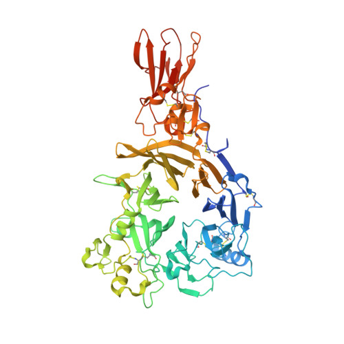
8RMJ - PubMed Abstract:
The precise assembly of a functional nervous system relies on axon guidance cues. Beyond engaging their cognate receptors and initiating signaling cascades that modulate cytoskeletal dynamics, guidance cues also bind components of the extracellular matrix, notably proteoglycans, yet the role and mechanisms of these interactions remain poorly understood. We found that Drosophila secreted semaphorins bind specifically to glycosaminoglycan (GAG) chains of proteoglycans, showing a preference based on the degree of sulfation. Structural analysis of Sema2b unveiled multiple GAG-binding sites positioned outside canonical plexin-binding site, with the highest affinity binding site located at the C-terminal tail, characterized by a lysine-rich helical arrangement that appears to be conserved across secreted semaphorins. In vivo studies revealed a crucial role of the Sema2b C-terminal tail in specifying the trajectory of olfactory receptor neurons. We propose that secreted semaphorins tether to the cell surface through interactions with GAG chains of proteoglycans, facilitating their presentation to cognate receptors on passing axons.
- Department of Cell Biology, Faculty of Science, Charles University, Prague 128 43, Czechia.
Organizational Affiliation: