8RM6 | pdb_00008rm6

Crystal Structure of Human Androgen Receptor DNA Binding Domain Bound to its Response Element: C3(1)ARE


Experimental Data Snapshot

  • Method: X-RAY DIFFRACTION
  • Resolution: 2.05 Å
  • R-Value Free: 
    0.247 (Depositor), 0.249 (DCC) 
  • R-Value Work: 
    0.221 (Depositor), 0.223 (DCC) 
  • R-Value Observed: 
    0.223 (Depositor) 

Starting Model: experimental
View more details

wwPDB Validation 3D Report Full Report

Validation slider image for 8RM6

This is version 1.0 of the entry. See complete history

Literature

Structural mechanism underlying variations in DNA binding by the androgen receptor.

Lee, X.Y.Van Eynde, W.Helsen, C.Willems, H.Peperstraete, K.De Block, S.Voet, A.Claessens, F.

(2024) J Steroid Biochem Mol Biol 241: 106499-106499

  • DOI: https://doi.org/10.1016/j.jsbmb.2024.106499
  • Primary Citation Related Structures: 
    8RM6, 8RM7

  • PubMed Abstract: 

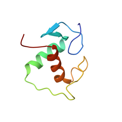
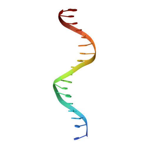
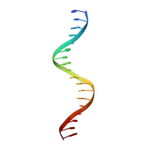
    The androgen receptor (AR) is a steroid activated transcription factor which recognizes DNA motifs resembling inverted repeats of a conserved 5'-AGAACA-3'-like hexanucleotides separated by a three-nucleotide spacer from a similar, but less conserved hexanucleotide. Here, we report the structures of the human AR DNA binding domain (DBD) bound to two natural AREs (C3 and MTV) in head-to-head dimer conformations, diffracting at 2.05 Å and 2.25 Å, respectively. These structures help to explain the impact of androgen insensitivity mutations on the structure integrity, DNA binding and DBD dimerization. The binding affinity of the AR DBD to different DNA motifs were measured by the BioLayer Interferometry (BLI) and further validated by Molecular Dynamics (MD) simulations. This shows that the high binding affinity of the first DBD to the upstream 5'-AGAACA-3' motif induces the cooperative binding of the second DBD to the second hexanucleotide. Our data indicate identical interaction of the DBDs to the upstream hexanucleotides, while forming an induced closer contact of the second DBD on the non-canonical hexanucleotides. The variation in binding between the DBD monomers are the result of differences in DNA occupancy, protein-protein interactions, DNA binding affinity, and DNA binding energy profiles. We propose this has functional consequences.


  • Organizational Affiliation
    • Molecular Endocrinology Laboratory, Department of Cellular and Molecular Medicine, Campus Gasthuisberg ON1 Herestraat 49 - box 901, Leuven 3000, Belgium.

Macromolecule Content 

  • Total Structure Weight: 27.38 kDa 
  • Atom Count: 1,908 
  • Modeled Residue Count: 182 
  • Deposited Residue Count: 182 
  • Unique protein chains: 1
  • Unique nucleic acid chains: 2

Macromolecules


Find similar proteins by:|  3D Structure
Entity ID: 1
MoleculeChains  Sequence LengthOrganismDetailsImage
Isoform 2 of Androgen receptorA [auth B],
B [auth A]
73Homo sapiensMutation(s): 0 
Gene Names: ARDHTRNR3C4
UniProt & NIH Common Fund Data Resources
Find proteins for P10275 (Homo sapiens)
Explore P10275 
Go to UniProtKB:  P10275
PHAROS:  P10275
GTEx:  ENSG00000169083 
Entity Groups
Sequence Clusters30% Identity50% Identity70% Identity90% Identity95% Identity100% Identity
UniProt GroupP10275
Sequence Annotations
Expand
Reference Sequence
Find similar nucleic acids by:  Sequence
Entity ID: 2
MoleculeChains LengthOrganismImage
C3(1)ARE_Chain DC [auth D]18Homo sapiens
Sequence Annotations
Expand
Reference Sequence
Find similar nucleic acids by:  Sequence
Entity ID: 3
MoleculeChains LengthOrganismImage
C3(1)ARE_Chain CD [auth C]18Homo sapiens
Sequence Annotations
Expand
Reference Sequence

Experimental Data & Validation

Experimental Data

  • Method: X-RAY DIFFRACTION
  • Resolution: 2.05 Å
  • R-Value Free:  0.247 (Depositor), 0.249 (DCC) 
  • R-Value Work:  0.221 (Depositor), 0.223 (DCC) 
  • R-Value Observed: 0.223 (Depositor) 
Space Group: P 2 21 2
Unit Cell:
Length ( Å )Angle ( ˚ )
a = 37.029α = 90
b = 95.359β = 90
c = 115.353γ = 90
Software Package:
Software NamePurpose
PHENIXrefinement
Aimlessdata scaling
XDSdata reduction
PHASERphasing

Structure Validation

View Full Validation Report


Entry History 

& Funding Information

Deposition Data


Funding OrganizationLocationGrant Number
Research Foundation - Flanders (FWO)BelgiumKU Leuven grant (GOA/19/100)

Revision History  (Full details and data files)

  • Version 1.0: 2024-04-24
    Type: Initial release