8RKD | pdb_00008rkd

TadA/CpaF with AMPPNP


Experimental Data Snapshot

  • Method: ELECTRON MICROSCOPY
  • Resolution: 3.80 Å
  • Aggregation State: PARTICLE 
  • Reconstruction Method: SINGLE PARTICLE 

wwPDB Validation 3D Report Full Report

Validation slider image for 8RKD

This is version 1.0 of the entry. See complete history

Literature

Bidirectional pilus processing in the Tad pilus system motor CpaF.

Hohl, M.Banks, E.J.Manley, M.P.Le, T.B.K.Low, H.H.

(2024) Nat Commun 15: 6635-6635

  • DOI: https://doi.org/10.1038/s41467-024-50280-6
  • Primary Citation Related Structures: 
    8RJF, 8RKD, 8RKL

  • PubMed Abstract: 

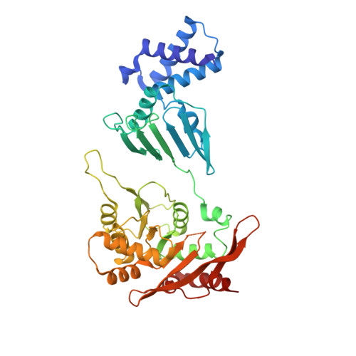
    The bacterial tight adherence pilus system (TadPS) assembles surface pili essential for adhesion and colonisation in many human pathogens. Pilus dynamics are powered by the ATPase CpaF (TadA), which drives extension and retraction cycles in Caulobacter crescentus through an unknown mechanism. Here we use cryogenic electron microscopy and cell-based light microscopy to characterise CpaF mechanism. We show that CpaF assembles into a hexamer with C2 symmetry in different nucleotide states. Nucleotide cycling occurs through an intra-subunit clamp-like mechanism that promotes sequential conformational changes between subunits. Moreover, a comparison of the active sites with different nucleotides bound suggests a mechanism for bidirectional motion. Conserved CpaF residues, predicted to interact with platform proteins CpaG (TadB) and CpaH (TadC), are mutated in vivo to establish their role in pilus processing. Our findings provide a model for how CpaF drives TadPS pilus dynamics and have broad implications for how other ancient type 4 filament family members power pilus assembly.


  • Organizational Affiliation
    • Department of Infectious Disease, Imperial College, London, UK.

Macromolecule Content 

  • Total Structure Weight: 282.51 kDa 
  • Atom Count: 18,616 
  • Modeled Residue Count: 2,514 
  • Deposited Residue Count: 2,538 
  • Unique protein chains: 1

Macromolecules

Find similar proteins by:|  3D Structure
Entity ID: 1
MoleculeChains  Sequence LengthOrganismDetailsImage
Pilus assembly ATPase CpaF423Caulobacter vibrioides NA1000Mutation(s): 0 
Gene Names: cpaFCCNA_03037
UniProt
Find proteins for A0A0H3CDS2 (Caulobacter vibrioides (strain NA1000 / CB15N))
Explore A0A0H3CDS2 
Go to UniProtKB:  A0A0H3CDS2
Entity Groups
Sequence Clusters30% Identity50% Identity70% Identity90% Identity95% Identity100% Identity
UniProt GroupA0A0H3CDS2
Sequence Annotations
Expand
Reference Sequence

Small Molecules

Ligands 4 Unique
IDChains Name / Formula / InChI Key2D Diagram3D Interactions
ANP

Query on ANP


Download:Ideal Coordinates CCD File
L [auth A],
R [auth D]
PHOSPHOAMINOPHOSPHONIC ACID-ADENYLATE ESTER
C10 H17 N6 O12 P3
PVKSNHVPLWYQGJ-KQYNXXCUSA-N
ADP

Query on ADP


Download:Ideal Coordinates CCD File
G [auth E],
J [auth F],
N [auth B],
P [auth C]
ADENOSINE-5'-DIPHOSPHATE
C10 H15 N5 O10 P2
XTWYTFMLZFPYCI-KQYNXXCUSA-N
EPE

Query on EPE


Download:Ideal Coordinates CCD File
H [auth E],
I [auth F],
M [auth B],
O [auth C]
4-(2-HYDROXYETHYL)-1-PIPERAZINE ETHANESULFONIC ACID
C8 H18 N2 O4 S
JKMHFZQWWAIEOD-UHFFFAOYSA-N
MG

Query on MG


Download:Ideal Coordinates CCD File
K [auth F],
Q [auth C]
MAGNESIUM ION
Mg
JLVVSXFLKOJNIY-UHFFFAOYSA-N

Experimental Data & Validation

Experimental Data

  • Method: ELECTRON MICROSCOPY
  • Resolution: 3.80 Å
  • Aggregation State: PARTICLE 
  • Reconstruction Method: SINGLE PARTICLE 
EM Software:
TaskSoftware PackageVersion
MODEL REFINEMENTPHENIX1.21rc1_4933:

Structure Validation

View Full Validation Report


Entry History 

& Funding Information

Deposition Data


Funding OrganizationLocationGrant Number
Wellcome TrustUnited Kingdom--

Revision History  (Full details and data files)

  • Version 1.0: 2024-08-14
    Type: Initial release