8RK5

Tail fibres of bacteriophage JBD30

  • Classification: VIRUS
  • Organism(s): Pseudomonas phage JBD30
  • Mutation(s): No 

  • Deposited: 2023-12-23 Released: 2024-08-14 
  • Deposition Author(s): Valentova, L., Fuzik, T., Plevka, P.
  • Funding Organization(s): Ministry of Education, Youth and Sports of the Czech Republic, European Research Council (ERC)

Experimental Data Snapshot

  • Method: ELECTRON MICROSCOPY
  • Resolution: 4.54 Å
  • Aggregation State: PARTICLE 
  • Reconstruction Method: SINGLE PARTICLE 

wwPDB Validation   3D Report Full Report


This is version 1.1 of the entry. See complete history


Literature

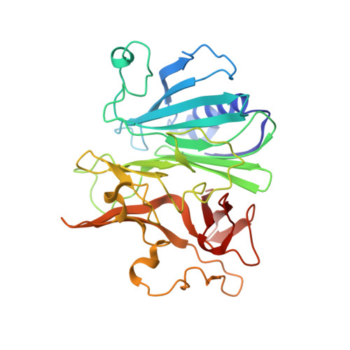
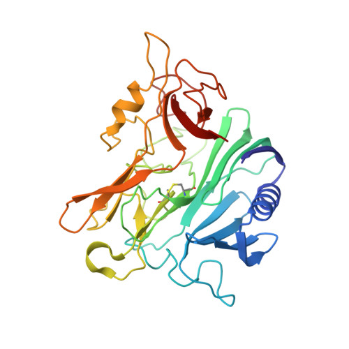
Structure and replication of Pseudomonas aeruginosa phage JBD30.

Valentova, L.Fuzik, T.Novacek, J.Hlavenkova, Z.Pospisil, J.Plevka, P.

(2024) EMBO J 

  • DOI: https://doi.org/10.1038/s44318-024-00195-1
  • Primary Citation of Related Structures:  
    8RK3, 8RK4, 8RK5, 8RK6, 8RK7, 8RK8, 8RK9, 8RKA, 8RKB, 8RKC, 8RKN, 8RKO, 8RKX, 8RQE

  • PubMed Abstract: 

    Bacteriophages are the most abundant biological entities on Earth, but our understanding of many aspects of their lifecycles is still incomplete. Here, we have structurally analysed the infection cycle of the siphophage Casadabanvirus JBD30. Using its baseplate, JBD30 attaches to Pseudomonas aeruginosa via the bacterial type IV pilus, whose subsequent retraction brings the phage to the bacterial cell surface. Cryo-electron microscopy structures of the baseplate-pilus complex show that the tripod of baseplate receptor-binding proteins attaches to the outer bacterial membrane. The tripod and baseplate then open to release three copies of the tape-measure protein, an event that is followed by DNA ejection. JBD30 major capsid proteins assemble into procapsids, which expand by 7% in diameter upon filling with phage dsDNA. The DNA-filled heads are finally joined with 180-nm-long tails, which bend easily because flexible loops mediate contacts between the successive discs of major tail proteins. It is likely that the structural features and replication mechanisms described here are conserved among siphophages that utilize the type IV pili for initial cell attachment.


  • Organizational Affiliation

    Central European Institute of Technology, Masaryk University, Brno, Czech Republic.


Macromolecules
Find similar proteins by:  (by identity cutoff)  |  3D Structure
Entity ID: 1
MoleculeChains Sequence LengthOrganismDetailsImage
Virion structural proteinA,
C,
E [auth D]
318Pseudomonas phage JBD30Mutation(s): 0 
UniProt
Find proteins for L7P7R6 (Pseudomonas phage JBD30)
Explore L7P7R6 
Go to UniProtKB:  L7P7R6
Entity Groups  
Sequence Clusters30% Identity50% Identity70% Identity90% Identity95% Identity100% Identity
UniProt GroupL7P7R6
Sequence Annotations
Expand
  • Reference Sequence
Find similar proteins by:  (by identity cutoff)  |  3D Structure
Entity ID: 2
MoleculeChains Sequence LengthOrganismDetailsImage
Virion structural proteinB,
D [auth E],
F
307Pseudomonas phage JBD30Mutation(s): 0 
UniProt
Find proteins for L7P7X2 (Pseudomonas phage JBD30)
Explore L7P7X2 
Go to UniProtKB:  L7P7X2
Entity Groups  
Sequence Clusters30% Identity50% Identity70% Identity90% Identity95% Identity100% Identity
UniProt GroupL7P7X2
Sequence Annotations
Expand
  • Reference Sequence
Experimental Data & Validation

Experimental Data

  • Method: ELECTRON MICROSCOPY
  • Resolution: 4.54 Å
  • Aggregation State: PARTICLE 
  • Reconstruction Method: SINGLE PARTICLE 
EM Software:
TaskSoftware PackageVersion
RECONSTRUCTIONRELION3.1
MODEL REFINEMENTPHENIX1.20.1

Structure Validation

View Full Validation Report


Entry History & Funding Information

Deposition Data


Funding OrganizationLocationGrant Number
Ministry of Education, Youth and Sports of the Czech RepublicCzech RepublicLX22NPO5103
European Research Council (ERC)European Union101043452

Revision History  (Full details and data files)

  • Version 1.0: 2024-08-14
    Type: Initial release
  • Version 1.1: 2024-08-28
    Changes: Data collection, Database references