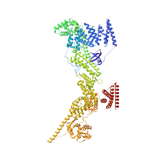
8RHZ

Structure of CUL9-RBX1 ubiquitin E3 ligase complex in unneddylated conformation - symmetry expanded unneddylated dimer


Experimental Data Snapshot

  • Method: ELECTRON MICROSCOPY
  • Resolution: 3.37 Å
  • Aggregation State: PARTICLE 
  • Reconstruction Method: SINGLE PARTICLE 

wwPDB Validation   3D Report Full Report


This is version 1.2 of the entry. See complete history


Literature

Noncanonical assembly, neddylation and chimeric cullin-RING/RBR ubiquitylation by the 1.8 MDa CUL9 E3 ligase complex.

Horn-Ghetko, D.Hopf, L.V.M.Tripathi-Giesgen, I.Du, J.Kostrhon, S.Vu, D.T.Beier, V.Steigenberger, B.Prabu, J.R.Stier, L.Bruss, E.M.Mann, M.Xiong, Y.Schulman, B.A.

(2024) Nat Struct Mol Biol 31: 1083-1094

  • DOI: https://doi.org/10.1038/s41594-024-01257-y
  • Primary Citation of Related Structures:  
    8Q7E, 8Q7H, 8RHZ

  • PubMed Abstract: 

    Ubiquitin ligation is typically executed by hallmark E3 catalytic domains. Two such domains, 'cullin-RING' and 'RBR', are individually found in several hundred human E3 ligases, and collaborate with E2 enzymes to catalyze ubiquitylation. However, the vertebrate-specific CUL9 complex with RBX1 (also called ROC1), of interest due to its tumor suppressive interaction with TP53, uniquely encompasses both cullin-RING and RBR domains. Here, cryo-EM, biochemistry and cellular assays elucidate a 1.8-MDa hexameric human CUL9-RBX1 assembly. Within one dimeric subcomplex, an E2-bound RBR domain is activated by neddylation of its own cullin domain and positioning from the adjacent CUL9-RBX1 in trans. Our data show CUL9 as unique among RBX1-bound cullins in dependence on the metazoan-specific UBE2F neddylation enzyme, while the RBR domain protects it from deneddylation. Substrates are recruited to various upstream domains, while ubiquitylation relies on both CUL9's neddylated cullin and RBR domains achieving self-assembled and chimeric cullin-RING/RBR E3 ligase activity.


  • Organizational Affiliation

    Department of Molecular Machines and Signaling, Max Planck Institute of Biochemistry, Martinsried, Germany.


Macromolecules
Find similar proteins by:  (by identity cutoff)  |  3D Structure
Entity ID: 1
MoleculeChains Sequence LengthOrganismDetailsImage
Cullin-9A,
C [auth B]
2,519Homo sapiensMutation(s): 0 
Gene Names: CUL9H7AP1KIAA0708PARC
UniProt & NIH Common Fund Data Resources
Find proteins for Q8IWT3 (Homo sapiens)
Go to UniProtKB:  Q8IWT3
PHAROS:  Q8IWT3
GTEx:  ENSG00000112659 
Sequence Annotations
Expand
  • Reference Sequence
Find similar proteins by:  (by identity cutoff)  |  3D Structure
Entity ID: 2
MoleculeChains Sequence LengthOrganismDetailsImage
E3 ubiquitin-protein ligase RBX1B [auth C],
D
108Homo sapiensMutation(s): 0 
Gene Names: RBX1RNF75ROC1
EC: 2.3.2.27 (PDB Primary Data), 2.3.2.32 (PDB Primary Data)
UniProt & NIH Common Fund Data Resources
Find proteins for P62877 (Homo sapiens)
Go to UniProtKB:  P62877
PHAROS:  P62877
GTEx:  ENSG00000100387 
Sequence Annotations
Expand
  • Reference Sequence
Experimental Data & Validation

Experimental Data

  • Method: ELECTRON MICROSCOPY
  • Resolution: 3.37 Å
  • Aggregation State: PARTICLE 
  • Reconstruction Method: SINGLE PARTICLE 
EM Software:
TaskSoftware PackageVersion
MODEL REFINEMENTPHENIX1.17.1_3660:

Structure Validation

View Full Validation Report


Entry History & Funding Information

Deposition Data


Funding OrganizationLocationGrant Number
European Research Council (ERC)European Union789016
Max Planck SocietyGermany--

Revision History  (Full details and data files)

  • Version 1.0: 2024-04-17
    Type: Initial release
  • Version 1.1: 2024-04-24
    Changes: Database references
  • Version 1.2: 2024-07-31
    Changes: Data collection, Database references