Structural dynamics and functional cooperativity of human NQO1 by ambient temperature serial crystallography and simulations.
Grieco, A., Boneta, S., Gavira, J.A., Pey, A.L., Basu, S., Orlans, J., Sanctis, D., Medina, M., Martin-Garcia, J.M.(2024) Protein Sci 33: e4957-e4957
- PubMed: 38501509 Search on PubMedSearch on PubMed Central
- DOI: https://doi.org/10.1002/pro.4957
- Primary Citation Related Structures:
8RFM, 8RFN - PubMed Abstract:
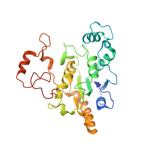
The human NQO1 (hNQO1) is a flavin adenine nucleotide (FAD)-dependent oxidoreductase that catalyzes the two-electron reduction of quinones to hydroquinones, being essential for the antioxidant defense system, stabilization of tumor suppressors, and activation of quinone-based chemotherapeutics. Moreover, it is overexpressed in several tumors, which makes it an attractive cancer drug target. To decipher new structural insights into the flavin reductive half-reaction of the catalytic mechanism of hNQO1, we have carried serial crystallography experiments at new ID29 beamline of the ESRF to determine, to the best of our knowledge, the first structure of the hNQO1 in complex with NADH. We have also performed molecular dynamics simulations of free hNQO1 and in complex with NADH. This is the first structural evidence that the hNQO1 functional cooperativity is driven by structural communication between the active sites through long-range propagation of cooperative effects across the hNQO1 structure. Both structural results and MD simulations have supported that the binding of NADH significantly decreases protein dynamics and stabilizes hNQO1 especially at the dimer core and interface. Altogether, these results pave the way for future time-resolved studies, both at x-ray free-electron lasers and synchrotrons, of the dynamics of hNQO1 upon binding to NADH as well as during the FAD cofactor reductive half-reaction. This knowledge will allow us to reveal unprecedented structural information of the relevance of the dynamics during the catalytic function of hNQO1.
- Department of Crystallography and Structural Biology, Institute of Physical Chemistry Blas Cabrera, Spanish National Research Council (CSIC), Madrid, Spain.
Organizational Affiliation: