


The crystal structure of a hydrated gramicidin S urea complex
Dodson, E.J.(1978) Nature 275: 206-207
Experimental Data Snapshot
wwPDB Validation 3D Report Full Report

Entity ID: 1 | |||||
|---|---|---|---|---|---|
| Molecule | Chains | Sequence Length | Organism | Details | Image |
| Gramicidin S | A [auth D] | 10 | Brevibacillus brevis | Mutation(s): 0 | 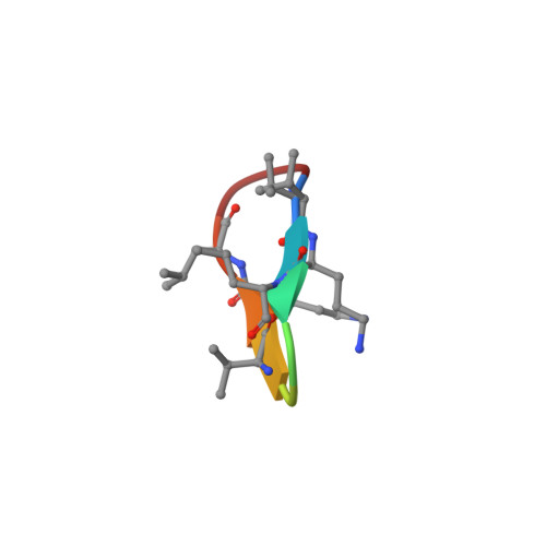 |
| Ligands 1 Unique | |||||
|---|---|---|---|---|---|
| ID | Chains | Name / Formula / InChI Key | 2D Diagram | 3D Interactions | |
| URE Download:Ideal Coordinates CCD File | B [auth D] | UREA C H4 N2 O XSQUKJJJFZCRTK-UHFFFAOYSA-N |  | ||
| Modified Residues 1 Unique | |||||
|---|---|---|---|---|---|
| ID | Chains | Type | Formula | 2D Diagram | Parent |
| DPN Query on DPN | A [auth D] | D-PEPTIDE LINKING | C9 H11 N O2 |  | -- |
| ORN Query on ORN | A [auth D] | L-PEPTIDE LINKING | C5 H12 N2 O2 |  | ALA |
| Entity ID: 1 | |||||
|---|---|---|---|---|---|
| ID | Chains | Name | Type/Class | 2D Diagram | 3D Interactions |
| PRD_000219 Query on PRD_000219 | A [auth D] | GRAMICIDIN S | Cyclic peptide / Antibiotic |  | |
| Length ( Å ) | Angle ( ˚ ) |
|---|---|
| a = 25.8 | α = 90 |
| b = 25.8 | β = 90 |
| c = 21.49 | γ = 120 |
| Software Name | Purpose |
|---|---|
| REFMAC | refinement |
| Funding Organization | Location | Grant Number |
|---|---|---|
| Not funded | -- |


