8RAX | pdb_00008rax

The crystal structure of DNA-bound human MutSbeta (MSH2_G674D/MSH3) in the canonical mismatch bound conformation, nucleotide free


Experimental Data Snapshot

  • Method: X-RAY DIFFRACTION
  • Resolution: 3.16 Å
  • R-Value Free: 
    0.292 (Depositor), 0.286 (DCC) 
  • R-Value Work: 
    0.257 (Depositor), 0.255 (DCC) 
  • R-Value Observed: 
    0.259 (Depositor) 

Starting Model: experimental
View more details

wwPDB Validation   3D Report Full Report


This is version 1.1 of the entry. See complete history


Literature

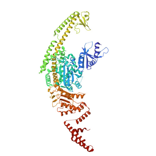
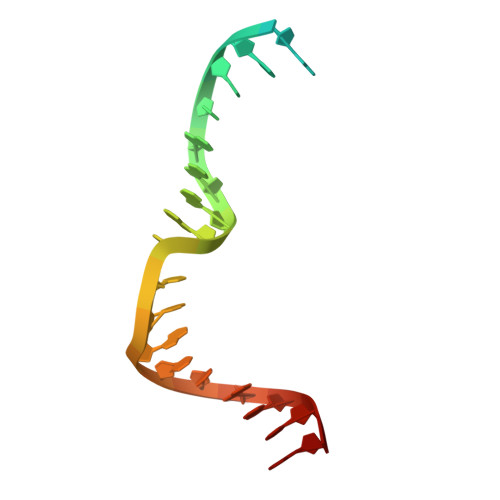
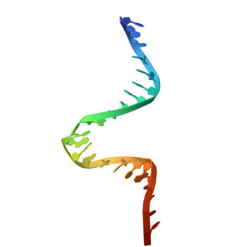
The crystal structure of DNA-bound human MutSbeta (MSH2/MSH3) in the canonical mismatch bound conformation with ADP bound in MSH2 and MSH3

Thomsen, M.Neudegger, T.Thieulin-Pardo, G.Blaesse, M.Costanzi, E.Steinbacher, S.Plotnikov, N.V.Dominguez, C.Iyer, R.R.Wilkinson, H.A.Monteagudo, E.Haque, T.S.Prasad, B.C.Finley, M.Boudet, J.Vogt, T.F.Felsenfeld, D.P.

To be published.

Macromolecules

Find similar proteins by:  (by identity cutoff)  |  3D Structure
Entity ID: 1
MoleculeChains Sequence LengthOrganismDetailsImage
DNA mismatch repair protein Msh2934Homo sapiensMutation(s): 1 
Gene Names: MSH2
UniProt & NIH Common Fund Data Resources
Find proteins for P43246 (Homo sapiens)
Explore P43246 
Go to UniProtKB:  P43246
PHAROS:  P43246
GTEx:  ENSG00000095002 
Entity Groups  
Sequence Clusters30% Identity50% Identity70% Identity90% Identity95% Identity100% Identity
UniProt GroupP43246
Sequence Annotations
Expand
  • Reference Sequence
Find similar proteins by:  (by identity cutoff)  |  3D Structure
Entity ID: 2
MoleculeChains Sequence LengthOrganismDetailsImage
DNA mismatch repair protein Msh3918Homo sapiensMutation(s): 0 
Gene Names: MSH3
UniProt & NIH Common Fund Data Resources
Find proteins for P20585 (Homo sapiens)
Explore P20585 
Go to UniProtKB:  P20585
PHAROS:  P20585
GTEx:  ENSG00000113318 
Entity Groups  
Sequence Clusters30% Identity50% Identity70% Identity90% Identity95% Identity100% Identity
UniProt GroupP20585
Sequence Annotations
Expand
  • Reference Sequence

Find similar nucleic acids by:  Sequence   |   3D Structure  

Entity ID: 3
MoleculeChains LengthOrganismImage
DNA (5'-D(P*AP*TP*CP*TP*GP*AP*AP*GP*CP*CP*GP*AP*TP*CP*GP*AP*TP*G)-3')24synthetic construct
Sequence Annotations
Expand
  • Reference Sequence

Find similar nucleic acids by:  Sequence   |   3D Structure  

Entity ID: 4
MoleculeChains LengthOrganismImage
DNA (5'-D(*TP*CP*AP*TP*CP*GP*AP*TP*CP*GP*CP*AP*GP*CP*TP*TP*CP*AP*GP*AP*TP*A)-3')24synthetic construct
Sequence Annotations
Expand
  • Reference Sequence
Experimental Data & Validation

Experimental Data

  • Method: X-RAY DIFFRACTION
  • Resolution: 3.16 Å
  • R-Value Free:  0.292 (Depositor), 0.286 (DCC) 
  • R-Value Work:  0.257 (Depositor), 0.255 (DCC) 
  • R-Value Observed: 0.259 (Depositor) 
Space Group: P 1
Unit Cell:
Length ( Å )Angle ( ˚ )
a = 66.899α = 67.68
b = 90.998β = 86.73
c = 96.162γ = 73.86
Software Package:
Software NamePurpose
REFMACrefinement
autoPROCdata reduction
autoPROCdata scaling
PHASERphasing

Structure Validation

View Full Validation Report


Entry History & Funding Information

Deposition Data


Funding OrganizationLocationGrant Number
CHDI FoundationUnited States--

Revision History  (Full details and data files)

  • Version 1.0: 2025-06-11
    Type: Initial release
  • Version 1.1: 2025-07-09
    Changes: Structure summary