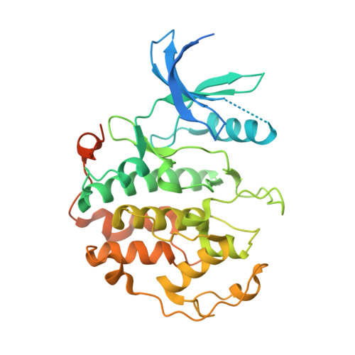
8R9A

A soakable crystal form of human CDK7 in complex with AMP-PNP


Experimental Data Snapshot

  • Method: X-RAY DIFFRACTION
  • Resolution: 1.71 Å
  • R-Value Free: 0.294 
  • R-Value Work: 0.220 
  • R-Value Observed: 0.224 

wwPDB Validation   3D Report Full Report


Ligand Structure Quality Assessment 


This is version 1.2 of the entry. See complete history


Literature

Protein engineering enables a soakable crystal form of human CDK7 primed for high-throughput crystallography and structure-based drug design.

Mukherjee, M.Day, P.J.Laverty, D.Bueren-Calabuig, J.A.Woodhead, A.J.Griffiths-Jones, C.Hiscock, S.East, C.Boyd, S.O'Reilly, M.

(2024) Structure 32: 1040-1048.e3

  • DOI: https://doi.org/10.1016/j.str.2024.05.011
  • Primary Citation of Related Structures:  
    8R99, 8R9A, 8R9B, 8R9O, 8R9S, 8R9U, 8RU8

  • PubMed Abstract: 

    Cyclin dependent kinase 7 (CDK7) is an important therapeutic kinase best known for its dual role in cell cycle regulation and gene transcription. Here, we describe the application of protein engineering to generate constructs leading to high resolution crystal structures of human CDK7 in both active and inactive conformations. The active state of the kinase was crystallized by incorporation of an additional surface residue mutation (W132R) onto the double phosphomimetic mutant background (S164D and T170E) that yielded the inactive kinase structure. A novel back-soaking approach was developed to determine crystal structures of several clinical and pre-clinical inhibitors of this kinase, demonstrating the potential utility of the crystal system for structure-based drug design (SBDD). The crystal structures help to rationalize the mode of inhibition and the ligand selectivity profiles versus key anti-targets. The protein engineering approach described here illustrates a generally applicable strategy for structural enablement of challenging molecular targets.


  • Organizational Affiliation

    Astex Pharmaceuticals, Cambridge CB4 0QA, UK. Electronic address: manjeet.mukherjee@astx.com.


Macromolecules
Find similar proteins by:  (by identity cutoff)  |  3D Structure
Entity ID: 1
MoleculeChains Sequence LengthOrganismDetailsImage
Cyclin-dependent kinase 7371Homo sapiensMutation(s): 3 
Gene Names: CDK7
EC: 2.7.11.23 (UniProt), 2.7.11.22 (UniProt)
UniProt & NIH Common Fund Data Resources
Find proteins for P50613 (Homo sapiens)
Explore P50613 
Go to UniProtKB:  P50613
PHAROS:  P50613
GTEx:  ENSG00000134058 
Entity Groups  
Sequence Clusters30% Identity50% Identity70% Identity90% Identity95% Identity100% Identity
UniProt GroupP50613
Sequence Annotations
Expand
  • Reference Sequence
Small Molecules
Ligands 2 Unique
IDChains Name / Formula / InChI Key2D Diagram3D Interactions
I74 (Subject of Investigation/LOI)
Query on I74

Download Ideal Coordinates CCD File 
C [auth A](3R,4R)-4-[[[7-[(phenylmethyl)amino]-3-propan-2-yl-pyrazolo[1,5-a]pyrimidin-5-yl]amino]methyl]piperidin-3-ol
C22 H30 N6 O
YCVGLKWJKIKVBI-MJGOQNOKSA-N
PEG
Query on PEG

Download Ideal Coordinates CCD File 
B [auth A]DI(HYDROXYETHYL)ETHER
C4 H10 O3
MTHSVFCYNBDYFN-UHFFFAOYSA-N
Experimental Data & Validation

Experimental Data

  • Method: X-RAY DIFFRACTION
  • Resolution: 1.71 Å
  • R-Value Free: 0.294 
  • R-Value Work: 0.220 
  • R-Value Observed: 0.224 
  • Space Group: P 21 21 2
Unit Cell:
Length ( Å )Angle ( ˚ )
a = 144.433α = 90
b = 40.417β = 90
c = 68.676γ = 90
Software Package:
Software NamePurpose
REFMACrefinement
PDB_EXTRACTdata extraction
XDSdata reduction
Aimlessdata scaling
REFMACphasing

Structure Validation

View Full Validation Report


Ligand Structure Quality Assessment 


Entry History & Funding Information

Deposition Data


Funding OrganizationLocationGrant Number
Not funded--

Revision History  (Full details and data files)

  • Version 1.0: 2024-05-29
    Type: Initial release
  • Version 1.1: 2024-06-26
    Changes: Database references
  • Version 1.2: 2024-08-21
    Changes: Database references