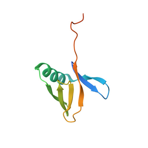
8R7O

HUWE1 WWE domain in complex with 2'F-ATP


Experimental Data Snapshot

  • Method: X-RAY DIFFRACTION
  • Resolution: 1.36 Å
  • R-Value Free: 0.195 
  • R-Value Work: 0.168 
  • R-Value Observed: 0.169 

Starting Model: experimental
View more details

wwPDB Validation   3D Report Full Report


Ligand Structure Quality Assessment 


This is version 1.1 of the entry. See complete history


Literature

A ligand discovery toolbox for the WWE domain family of human E3 ligases.

Munzker, L.Kimani, S.W.Fowkes, M.M.Dong, A.Zheng, H.Li, Y.Dasovich, M.Zak, K.M.Leung, A.K.L.Elkins, J.M.Kessler, D.Arrowsmith, C.H.Halabelian, L.Bottcher, J.

(2024) Commun Biol 7: 901-901

  • DOI: https://doi.org/10.1038/s42003-024-06584-w
  • Primary Citation of Related Structures:  
    8R5N, 8R6A, 8R6B, 8R7O, 8RD0, 8RD1, 8RD7

  • PubMed Abstract: 

    The WWE domain is a relatively under-researched domain found in twelve human proteins and characterized by a conserved tryptophan-tryptophan-glutamate (WWE) sequence motif. Six of these WWE domain-containing proteins also contain domains with E3 ubiquitin ligase activity. The general recognition of poly-ADP-ribosylated substrates by WWE domains suggests a potential avenue for development of Proteolysis-Targeting Chimeras (PROTACs). Here, we present novel crystal structures of the HUWE1, TRIP12, and DTX1 WWE domains in complex with PAR building blocks and their analogs, thus enabling a comprehensive analysis of the PAR binding site structural diversity. Furthermore, we introduce a versatile toolbox of biophysical and biochemical assays for the discovery and characterization of novel WWE domain binders, including fluorescence polarization-based PAR binding and displacement assays, 15 N-NMR-based binding affinity assays and 19 F-NMR-based competition assays. Through these assays, we have characterized the binding of monomeric iso-ADP-ribose (iso-ADPr) and its nucleotide analogs with the aforementioned WWE proteins. Finally, we have utilized the assay toolbox to screen a small molecule fragment library leading to the successful discovery of novel ligands targeting the HUWE1 WWE domain.


  • Organizational Affiliation

    Boehringer Ingelheim RCV GmbH & Co KG, Vienna, Austria.


Macromolecules
Find similar proteins by:  (by identity cutoff)  |  3D Structure
Entity ID: 1
MoleculeChains Sequence LengthOrganismDetailsImage
E3 ubiquitin-protein ligase HUWE1
A, B, C, D
91Homo sapiensMutation(s): 0 
Gene Names: HUWE1
EC: 2.3.2.26
UniProt & NIH Common Fund Data Resources
Find proteins for Q7Z6Z7 (Homo sapiens)
Explore Q7Z6Z7 
Go to UniProtKB:  Q7Z6Z7
PHAROS:  Q7Z6Z7
GTEx:  ENSG00000086758 
Entity Groups  
Sequence Clusters30% Identity50% Identity70% Identity90% Identity95% Identity100% Identity
UniProt GroupQ7Z6Z7
Sequence Annotations
Expand
  • Reference Sequence
Small Molecules
Ligands 3 Unique
IDChains Name / Formula / InChI Key2D Diagram3D Interactions
YGC (Subject of Investigation/LOI)
Query on YGC

Download Ideal Coordinates CCD File 
F [auth A],
I [auth C]
[[(2~{R},3~{R},4~{R},5~{R})-5-(6-aminopurin-9-yl)-4-fluoranyl-3-oxidanyl-oxolan-2-yl]methoxy-oxidanyl-phosphoryl] phosphono hydrogen phosphate
C10 H15 F N5 O12 P3
CCXCDDWASROAJA-QYYRPYCUSA-N
CL
Query on CL

Download Ideal Coordinates CCD File 
E [auth A],
G [auth B],
H [auth C]
CHLORIDE ION
Cl
VEXZGXHMUGYJMC-UHFFFAOYSA-M
NA
Query on NA

Download Ideal Coordinates CCD File 
J [auth C]SODIUM ION
Na
FKNQFGJONOIPTF-UHFFFAOYSA-N
Experimental Data & Validation

Experimental Data

  • Method: X-RAY DIFFRACTION
  • Resolution: 1.36 Å
  • R-Value Free: 0.195 
  • R-Value Work: 0.168 
  • R-Value Observed: 0.169 
  • Space Group: P 43 21 2
Unit Cell:
Length ( Å )Angle ( ˚ )
a = 62.915α = 90
b = 62.915β = 90
c = 231.945γ = 90
Software Package:
Software NamePurpose
PHENIXrefinement
XDSdata reduction
autoPROCdata scaling
PHASERphasing

Structure Validation

View Full Validation Report


Ligand Structure Quality Assessment 


Entry History & Funding Information

Deposition Data


Funding OrganizationLocationGrant Number
European Union (EU)European Union875510

Revision History  (Full details and data files)

  • Version 1.0: 2024-07-31
    Type: Initial release
  • Version 1.1: 2024-08-07
    Changes: Database references