8R6Y

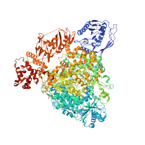
Structure of the SFTSV L protein stalled in a transcription-specific early elongation state with bound capped RNA [TRANSCRIPTION-EARLY-ELONGATION]


Experimental Data Snapshot

  • Method: ELECTRON MICROSCOPY
  • Resolution: 3.40 Å
  • Aggregation State: PARTICLE 
  • Reconstruction Method: SINGLE PARTICLE 

wwPDB Validation   3D Report Full Report


This is version 1.3 of the entry. See complete history


Literature

Structural snapshots of phenuivirus cap-snatching and transcription.

Williams, H.M.Thorkelsson, S.R.Vogel, D.Busch, C.Milewski, M.Cusack, S.Grunewald, K.Quemin, E.R.J.Rosenthal, M.

(2024) Nucleic Acids Res 52: 6049-6065

  • DOI: https://doi.org/10.1093/nar/gkae330
  • Primary Citation of Related Structures:  
    8R6U, 8R6W, 8R6Y

  • PubMed Abstract: 

    Severe fever with thrombocytopenia syndrome virus (SFTSV) is a human pathogen that is now endemic to several East Asian countries. The viral large (L) protein catalyzes viral transcription by stealing host mRNA caps via a process known as cap-snatching. Here, we establish an in vitro cap-snatching assay and present three high-quality electron cryo-microscopy (cryo-EM) structures of the SFTSV L protein in biologically relevant, transcription-specific states. In a priming-state structure, we show capped RNA bound to the L protein cap-binding domain (CBD). The L protein conformation in this priming structure is significantly different from published replication-state structures, in particular the N- and C-terminal domains. The capped-RNA is positioned in a way that it can feed directly into the RNA-dependent RNA polymerase (RdRp) ready for elongation. We also captured the L protein in an early-elongation state following primer-incorporation demonstrating that this priming conformation is retained at least in the very early stages of primer extension. This structural data is complemented by in vitro biochemical and cell-based assays. Together, these insights further our mechanistic understanding of how SFTSV and other bunyaviruses incorporate stolen host mRNA fragments into their viral transcripts thereby allowing the virus to hijack host cell translation machinery.


  • Organizational Affiliation

    Bernhard Nocht Institute for Tropical Medicine (BNITM), Hamburg, Germany.


Macromolecules

Find similar proteins by:  (by identity cutoff)  |  3D Structure
Entity ID: 1
MoleculeChains Sequence LengthOrganismDetailsImage
RNA-directed RNA polymerase L2,084SFTS virus AH12Mutation(s): 1 
EC: 3.1 (UniProt), 2.7.7.48 (UniProt)
UniProt
Find proteins for I0DF35 (Dabie bandavirus)
Explore I0DF35 
Go to UniProtKB:  I0DF35
Entity Groups  
Sequence Clusters30% Identity50% Identity70% Identity90% Identity95% Identity100% Identity
UniProt GroupI0DF35
Sequence Annotations
Expand
  • Reference Sequence

Find similar nucleic acids by:  Sequence   |   3D Structure  

Entity ID: 2
MoleculeChains LengthOrganismImage
RNA (5'-R(*AP*CP*AP*CP*AP*GP*AP*GP*AP*CP*GP*CP*CP*CP*AP*G)-3')B [auth P]20SFTS virus AH12
Sequence Annotations
Expand
  • Reference Sequence

Find similar nucleic acids by:  Sequence   |   3D Structure  

Entity ID: 3
MoleculeChains LengthOrganismImage
RNA (5'-R(P*CP*UP*GP*GP*GP*CP*GP*GP*UP*AP*AP*AP*UP*GP*U)-3')C [auth T]20SFTS virus AH12
Sequence Annotations
Expand
  • Reference Sequence

Find similar nucleic acids by:  Sequence   |   3D Structure  

Entity ID: 4
MoleculeChains LengthOrganismImage
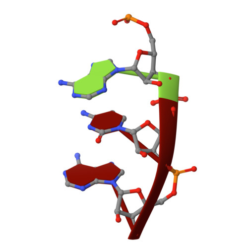
RNA (5'-R(P*AP*CP*A)-3')D [auth G]3SFTS virus AH12
Sequence Annotations
Expand
  • Reference Sequence

Find similar nucleic acids by:  Sequence   |   3D Structure  

Entity ID: 5
MoleculeChains LengthOrganismImage
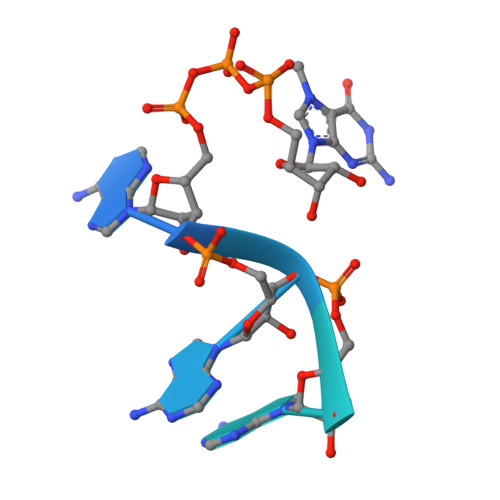
RNA (5'-R(*(M7G)*AP*AP*A)-3')E [auth C]18SFTS virus AH12
Sequence Annotations
Expand
  • Reference Sequence
Small Molecules
Ligands 2 Unique
IDChains Name / Formula / InChI Key2D Diagram3D Interactions
2KH (Subject of Investigation/LOI)
Query on 2KH

Download Ideal Coordinates CCD File 
F [auth A]5'-O-[(S)-hydroxy{[(S)-hydroxy(phosphonooxy)phosphoryl]amino}phosphoryl]uridine
C9 H16 N3 O14 P3
OZIBFYOFLVBDIY-XVFCMESISA-N
MG
Query on MG

Download Ideal Coordinates CCD File 
G [auth A]MAGNESIUM ION
Mg
JLVVSXFLKOJNIY-UHFFFAOYSA-N
Experimental Data & Validation

Experimental Data

  • Method: ELECTRON MICROSCOPY
  • Resolution: 3.40 Å
  • Aggregation State: PARTICLE 
  • Reconstruction Method: SINGLE PARTICLE 
EM Software:
TaskSoftware PackageVersion
MODEL REFINEMENTPHENIX
RECONSTRUCTIONRELION

Structure Validation

View Full Validation Report


Entry History & Funding Information

Deposition Data


Funding OrganizationLocationGrant Number
Leibniz AssociationGermanyK72/2017
German Research Foundation (DFG)GermanyNST 152/772-1, 774-1, 775-1 and 776-1
German Federal Ministry for Education and ResearchGermany01KI2019

Revision History  (Full details and data files)

  • Version 1.0: 2024-04-24
    Type: Initial release
  • Version 1.1: 2024-05-01
    Changes: Database references
  • Version 1.2: 2024-05-15
    Changes: Database references
  • Version 1.3: 2024-06-19
    Changes: Database references