8R50 | pdb_00008r50

Mouse teneurin-3 compact dimer - A1B1 isoform


Experimental Data Snapshot

  • Method: ELECTRON MICROSCOPY
  • Resolution: 3.10 Å
  • Aggregation State: PARTICLE 
  • Reconstruction Method: SINGLE PARTICLE 

wwPDB Validation 3D Report Full Report

Validation slider image for 8R50

This is version 1.2 of the entry. See complete history

Literature

Alternative splicing controls teneurin-3 compact dimer formation for neuronal recognition.

Gogou, C.Beugelink, J.W.Frias, C.P.Kresik, L.Jaroszynska, N.Drescher, U.Janssen, B.J.C.Hindges, R.Meijer, D.H.

(2024) Nat Commun 15: 3648-3648

  • DOI: https://doi.org/10.1038/s41467-024-47763-x
  • Primary Citation Related Structures: 
    8R50, 8R51, 8R54

  • PubMed Abstract: 

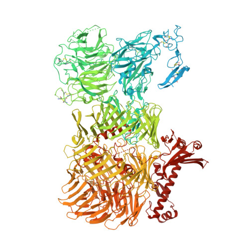
    Neuronal network formation is facilitated by recognition between synaptic cell adhesion molecules at the cell surface. Alternative splicing of cell adhesion molecules provides additional specificity in forming neuronal connections. For the teneurin family of cell adhesion molecules, alternative splicing of the EGF-repeats and NHL domain controls synaptic protein-protein interactions. Here we present cryo-EM structures of the compact dimeric ectodomain of two teneurin-3 isoforms that harbour the splice insert in the EGF-repeats. This dimer is stabilised by an EGF8-ABD contact between subunits. Cryo-EM reconstructions of all four splice variants, together with SAXS and negative stain EM, reveal compacted dimers for each, with variant-specific dimeric arrangements. This results in specific trans-cellular interactions, as tested in cell clustering and stripe assays. The compact conformations provide a structural basis for teneurin homo- and heterophilic interactions. Altogether, our findings demonstrate how alternative splicing results in rearrangements of the dimeric subunits, influencing neuronal recognition and likely circuit wiring.


  • Organizational Affiliation
    • Department of Bionanoscience, Kavli Institute of Nanoscience Delft, Delft University of Technology, van der Maasweg 9, Delft, the Netherlands.

Macromolecule Content 

  • Total Structure Weight: 544.38 kDa 
  • Atom Count: 29,752 
  • Modeled Residue Count: 3,778 
  • Deposited Residue Count: 4,814 
  • Unique protein chains: 1

Macromolecules

Find similar proteins by:|  3D Structure
Entity ID: 1
MoleculeChains  Sequence LengthOrganismDetailsImage
Teneurin-3A [auth B],
B [auth A]
2,407Mus musculusMutation(s): 0 
Gene Names: Tenm3Kiaa1455Odz3Tnm3
UniProt
Find proteins for Q9WTS6 (Mus musculus)
Explore Q9WTS6 
Go to UniProtKB:  Q9WTS6
Entity Groups
Sequence Clusters30% Identity50% Identity70% Identity90% Identity95% Identity100% Identity
UniProt GroupQ9WTS6
Glycosylation
Glycosylation Sites: 11Go to GlyGen: Q9WTS6-1
Sequence Annotations
Expand
Reference Sequence

Oligosaccharides

Help  
Entity ID: 2
MoleculeChains Length2D Diagram GlycosylationD Interactions
2-acetamido-2-deoxy-beta-D-glucopyranose-(1-4)-2-acetamido-2-deoxy-beta-D-glucopyranose
C, D, E, F, G
C, D, E, F, G, H, I, J
2N-Glycosylation
Glycosylation Resources
GlyTouCan: G42666HT
GlyCosmos: G42666HT
GlyGen: G42666HT

Small Molecules

Ligands 1 Unique
IDChains Name / Formula / InChI Key2D Diagram3D Interactions
NAG

Query on NAG


Download:Ideal Coordinates CCD File
K [auth B]
L [auth B]
M [auth B]
N [auth B]
O [auth B]
K [auth B],
L [auth B],
M [auth B],
N [auth B],
O [auth B],
P [auth B],
Q [auth B],
R [auth B],
S [auth A],
T [auth A],
U [auth A],
V [auth A],
W [auth A],
X [auth A],
Y [auth A],
Z [auth A]
2-acetamido-2-deoxy-beta-D-glucopyranose
C8 H15 N O6
OVRNDRQMDRJTHS-FMDGEEDCSA-N

Experimental Data & Validation

Experimental Data

  • Method: ELECTRON MICROSCOPY
  • Resolution: 3.10 Å
  • Aggregation State: PARTICLE 
  • Reconstruction Method: SINGLE PARTICLE 
EM Software:
TaskSoftware PackageVersion
MODEL REFINEMENTPHENIX

Structure Validation

View Full Validation Report


Entry History 

& Funding Information

Deposition Data


Funding OrganizationLocationGrant Number
Netherlands Organisation for Scientific Research (NWO)Netherlands722.016.004

Revision History  (Full details and data files)

  • Version 1.0: 2024-05-08
    Type: Initial release
  • Version 1.1: 2024-11-20
    Changes: Data collection, Structure summary
  • Version 1.2: 2025-07-09
    Changes: Data collection