8R1P

Pim1 in complex with 6-bromobenzo[d]oxazol-2-amine and Pimtide


Experimental Data Snapshot

  • Method: X-RAY DIFFRACTION
  • Resolution: 2.45 Å
  • R-Value Free: 0.206 
  • R-Value Work: 0.176 
  • R-Value Observed: 0.178 

wwPDB Validation   3D Report Full Report


Ligand Structure Quality Assessment 


This is version 1.1 of the entry. See complete history


Literature

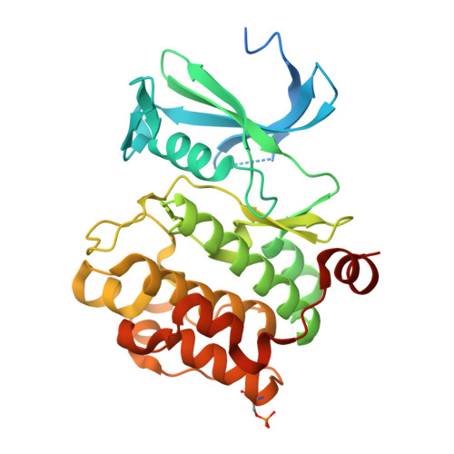
What doesn't fit is made to fit: Pim-1 kinase adapts to the configuration of stilbene-based inhibitors.

Hochban, P.M.M.Heyder, L.Heine, A.Diederich, W.E.

(2024) Arch Pharm (Weinheim) : e2400094-e2400094

  • DOI: https://doi.org/10.1002/ardp.202400094
  • Primary Citation of Related Structures:  
    8R10, 8R18, 8R1N, 8R1P, 8R1T

  • PubMed Abstract: 

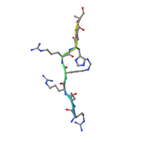
    Recently, we have developed novel Pim-1 kinase inhibitors starting from a dihydrobenzofuran core structure using a computational approach. Here, we report the design and synthesis of stilbene-based Pim-1 kinase inhibitors obtained by formal elimination of the dihydrofuran ring. These inhibitors of the first design cycle, which were obtained as inseparable cis/trans mixtures, showed affinities in the low single-digit micromolar range. To be able to further optimize these compounds in a structure-based fashion, we determined the X-ray structures of the protein-ligand-complexes. Surprisingly, only the cis-isomer binds upon crystallization of the cis/trans-mixture of the ligands with Pim-1 kinase and the substrate PIMTIDE, the binding mode being largely consistent with that predicted by docking. After crystallization of the exclusively trans-configured derivatives, a markedly different binding mode for the inhibitor and a concomitant rearrangement of the glycine-rich loop is observed, resulting in the ligand being deeply buried in the binding pocket.


  • Organizational Affiliation

    Institut für Pharmazeutische Chemie, Zentrum für Tumor und Immunbiologie, Philipps-Universität Marburg, Marburg, Germany.


Macromolecules
Find similar proteins by:  (by identity cutoff)  |  3D Structure
Entity ID: 1
MoleculeChains Sequence LengthOrganismDetailsImage
Serine/threonine-protein kinase pim-1313Homo sapiensMutation(s): 1 
Gene Names: PIM1
EC: 2.7.11.1
UniProt & NIH Common Fund Data Resources
Find proteins for P11309 (Homo sapiens)
Explore P11309 
Go to UniProtKB:  P11309
PHAROS:  P11309
GTEx:  ENSG00000137193 
Entity Groups  
Sequence Clusters30% Identity50% Identity70% Identity90% Identity95% Identity100% Identity
UniProt GroupP11309
Sequence Annotations
Expand
  • Reference Sequence

Find similar proteins by:  Sequence   |   3D Structure  

Entity ID: 2
MoleculeChains Sequence LengthOrganismDetailsImage
PimtideB [auth C]14Homo sapiensMutation(s): 0 
Entity Groups  
Sequence Clusters30% Identity50% Identity70% Identity90% Identity95% Identity100% Identity
Sequence Annotations
Expand
  • Reference Sequence
Small Molecules
Ligands 1 Unique
IDChains Name / Formula / InChI Key2D Diagram3D Interactions
XIP (Subject of Investigation/LOI)
Query on XIP

Download Ideal Coordinates CCD File 
C [auth A]4-(4-aminostyryl)benzoic acid
C15 H13 N O2
XLNJCEOZYNLROH-UHFFFAOYSA-N
Modified Residues  1 Unique
IDChains TypeFormula2D DiagramParent
SEP
Query on SEP
A
L-PEPTIDE LINKINGC3 H8 N O6 PSER
Experimental Data & Validation

Experimental Data

  • Method: X-RAY DIFFRACTION
  • Resolution: 2.45 Å
  • R-Value Free: 0.206 
  • R-Value Work: 0.176 
  • R-Value Observed: 0.178 
  • Space Group: P 65
Unit Cell:
Length ( Å )Angle ( ˚ )
a = 97.313α = 90
b = 97.313β = 90
c = 80.837γ = 120
Software Package:
Software NamePurpose
MxCuBEdata collection
PHENIXrefinement
XDSdata reduction
XDSdata scaling
PHASERphasing
Cootmodel building

Structure Validation

View Full Validation Report


Ligand Structure Quality Assessment 


Entry History & Funding Information

Deposition Data


Funding OrganizationLocationGrant Number
Not funded--

Revision History  (Full details and data files)

  • Version 1.0: 2024-03-20
    Type: Initial release
  • Version 1.1: 2024-05-22
    Changes: Database references