8R1L

Structure of avian H5N1 influenza A polymerase in complex with human ANP32B.


Experimental Data Snapshot

  • Method: ELECTRON MICROSCOPY
  • Resolution: 3.10 Å
  • Aggregation State: PARTICLE 
  • Reconstruction Method: SINGLE PARTICLE 

Starting Models: in silico, experimental
View more details

wwPDB Validation   3D Report Full Report


This is version 1.3 of the entry. See complete history


Literature

Structures of H5N1 influenza polymerase with ANP32B reveal mechanisms of genome replication and host adaptation.

Staller, E.Carrique, L.Swann, O.C.Fan, H.Keown, J.R.Sheppard, C.M.Barclay, W.S.Grimes, J.M.Fodor, E.

(2024) Nat Commun 15: 4123-4123

  • DOI: https://doi.org/10.1038/s41467-024-48470-3
  • Primary Citation of Related Structures:  
    8R1J, 8R1L

  • PubMed Abstract: 

    Avian influenza A viruses (IAVs) pose a public health threat, as they are capable of triggering pandemics by crossing species barriers. Replication of avian IAVs in mammalian cells is hindered by species-specific variation in acidic nuclear phosphoprotein 32 (ANP32) proteins, which are essential for viral RNA genome replication. Adaptive mutations enable the IAV RNA polymerase (FluPolA) to surmount this barrier. Here, we present cryo-electron microscopy structures of monomeric and dimeric avian H5N1 FluPolA with human ANP32B. ANP32B interacts with the PA subunit of FluPolA in the monomeric form, at the site used for its docking onto the C-terminal domain of host RNA polymerase II during viral transcription. ANP32B acts as a chaperone, guiding FluPolA towards a ribonucleoprotein-associated FluPolA to form an asymmetric dimer-the replication platform for the viral genome. These findings offer insights into the molecular mechanisms governing IAV genome replication, while enhancing our understanding of the molecular processes underpinning mammalian adaptations in avian-origin FluPolA.


  • Organizational Affiliation

    Sir William Dunn School of Pathology, University of Oxford, Oxford, UK.


Macromolecules
Find similar proteins by:  (by identity cutoff)  |  3D Structure
Entity ID: 1
MoleculeChains Sequence LengthOrganismDetailsImage
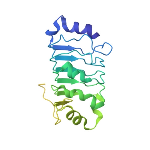
Acidic leucine-rich nuclear phosphoprotein 32 family member BA [auth D]251Homo sapiensMutation(s): 0 
Gene Names: ANP32B
UniProt & NIH Common Fund Data Resources
Find proteins for Q92688 (Homo sapiens)
Explore Q92688 
Go to UniProtKB:  Q92688
PHAROS:  Q92688
GTEx:  ENSG00000136938 
Entity Groups  
Sequence Clusters30% Identity50% Identity70% Identity90% Identity95% Identity100% Identity
UniProt GroupQ92688
Sequence Annotations
Expand
  • Reference Sequence
Find similar proteins by:  (by identity cutoff)  |  3D Structure
Entity ID: 2
MoleculeChains Sequence LengthOrganismDetailsImage
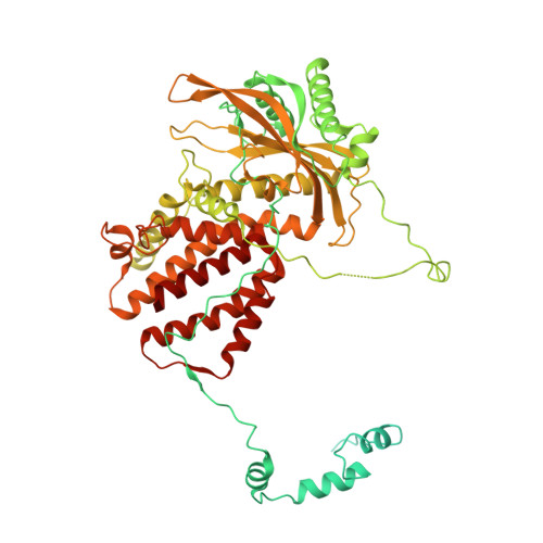
Polymerase acidic proteinB [auth A]716Influenza A virusMutation(s): 4 
Gene Names: PA
UniProt
Find proteins for A5Z236 (Influenza A virus)
Explore A5Z236 
Go to UniProtKB:  A5Z236
Entity Groups  
Sequence Clusters30% Identity50% Identity70% Identity90% Identity95% Identity100% Identity
UniProt GroupA5Z236
Sequence Annotations
Expand
  • Reference Sequence
Find similar proteins by:  (by identity cutoff)  |  3D Structure
Entity ID: 3
MoleculeChains Sequence LengthOrganismDetailsImage
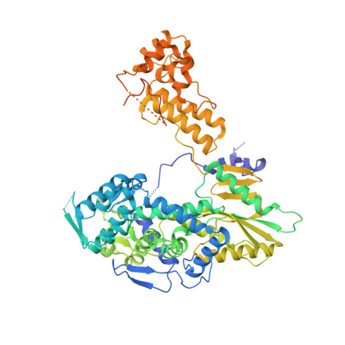
RNA-directed RNA polymerase catalytic subunitC [auth B]757Influenza A virusMutation(s): 4 
Gene Names: PB1
EC: 2.7.7.48
UniProt
Find proteins for A5Z231 (Influenza A virus)
Explore A5Z231 
Go to UniProtKB:  A5Z231
Entity Groups  
Sequence Clusters30% Identity50% Identity70% Identity90% Identity95% Identity100% Identity
UniProt GroupA5Z231
Sequence Annotations
Expand
  • Reference Sequence
Find similar proteins by:  (by identity cutoff)  |  3D Structure
Entity ID: 4
MoleculeChains Sequence LengthOrganismDetailsImage
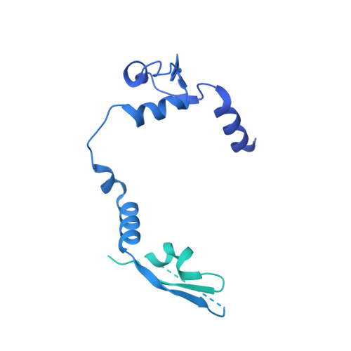
Polymerase basic protein 2D [auth C]898Influenza A virusMutation(s): 0 
Gene Names: PB2
UniProt
Find proteins for A5Z230 (Influenza A virus)
Explore A5Z230 
Go to UniProtKB:  A5Z230
Entity Groups  
Sequence Clusters30% Identity50% Identity70% Identity90% Identity95% Identity100% Identity
UniProt GroupA5Z230
Sequence Annotations
Expand
  • Reference Sequence
Experimental Data & Validation

Experimental Data

  • Method: ELECTRON MICROSCOPY
  • Resolution: 3.10 Å
  • Aggregation State: PARTICLE 
  • Reconstruction Method: SINGLE PARTICLE 
EM Software:
TaskSoftware PackageVersion
MODEL REFINEMENTCoot0.9.8.7
MODEL REFINEMENTPHENIX1.20
RECONSTRUCTIONcryoSPARCV4.2

Structure Validation

View Full Validation Report


Entry History & Funding Information

Deposition Data


Funding OrganizationLocationGrant Number
Wellcome TrustUnited Kingdom200835/Z/16/Z
Wellcome TrustUnited Kingdom222510/Z/21/Z
Medical Research Council (MRC, United Kingdom)United KingdomMR/R009945/1
Wellcome TrustUnited Kingdom093305/Z/10/Z
Wellcome TrustUnited Kingdom060208/Z/00/Z
Wellcome TrustUnited Kingdom203141/Z/16/Z

Revision History  (Full details and data files)

  • Version 1.0: 2024-05-08
    Type: Initial release
  • Version 1.1: 2024-05-29
    Changes: Database references
  • Version 1.2: 2024-07-24
    Changes: Data collection, Database references
  • Version 1.3: 2024-07-31
    Changes: Data collection, Database references