Primary Citation Related Structures: 8CIN, 8R1C, 8R1D
PubMed Abstract:
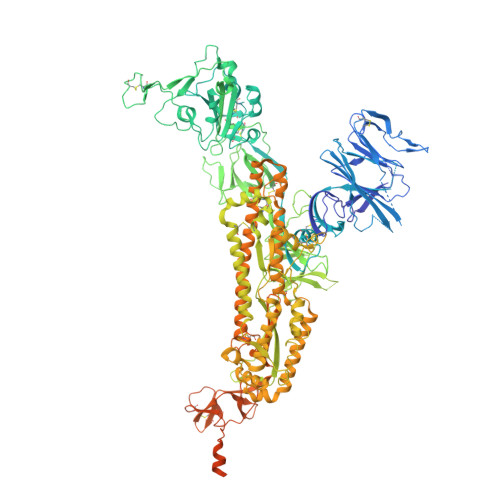
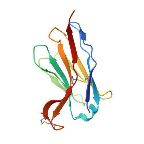
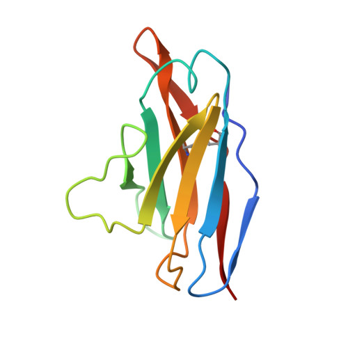
Under pressure from neutralising antibodies induced by vaccination or infection the SARS-CoV-2 spike gene has become a hotspot for evolutionary change, leading to the failure of all mAbs developed for clinical use. Most potent antibodies bind to the receptor binding domain which has become heavily mutated. Here we study responses to a conserved epitope in sub-domain-1 (SD1) of spike which have become more prominent because of mutational escape from antibodies directed to the receptor binding domain. Some SD1 reactive mAbs show potent and broad neutralization of SARS-CoV-2 variants. We structurally map the dominant SD1 epitope and provide a mechanism of action by blocking interaction with ACE2. Mutations in SD1 have not been sustained to date, but one, E554K, leads to escape from mAbs. This mutation has now emerged in several sublineages including BA.2.86, reflecting selection pressure on the virus exerted by the increasing prominence of the anti-SD1 response.
Organizational Affiliation:
Chinese Academy of Medical Science (CAMS) Oxford Institute (COI), University of Oxford, Oxford, UK.
Division of Structural Biology, Nuffield Department of Medicine, University of Oxford, Centre for Human Genetics, Oxford, UK.
College of Life Sciences, Zhejiang University, Hangzhou, 310058, China.
Centre for Human Genetics, Nuffield Department of Medicine, University of Oxford, Oxford, UK.
NIHR Oxford Biomedical Research Centre, Oxford University Hospitals NHS Foundation Trust, Oxford, UK.
Division of Emerging Infectious Disease, Research Department, Faculty of Medicine Siriraj Hospital, Mahidol University, Bangkok-Noi, Bangkok, 10700, Thailand.
Viral Pseudotype Unit, Medway School of Pharmacy, University of Kent and Greenwich Chatham Maritime, Kent, ME4 4TB, UK.
Peter Medawar Building for Pathogen Research, University of Oxford, Oxford, UK.
Translational Gastroenterology Unit, Nuffield Department of Medicine, University of Oxford, Oxford, UK.
NDM Centre For Global Health Research, Nuffield Department of Medicine, University of Oxford, Oxford, UK.
Mahidol-Oxford Tropical Medicine Research Unit, Bangkok, Thailand.
Division of Structural Biology, Nuffield Department of Medicine, University of Oxford, Centre for Human Genetics, Oxford, UK. liz@strubi.ox.ac.uk.
Chinese Academy of Medical Science (CAMS) Oxford Institute (COI), University of Oxford, Oxford, UK. juthathip.mongkolsapaya@well.ox.ac.uk.
Centre for Human Genetics, Nuffield Department of Medicine, University of Oxford, Oxford, UK. juthathip.mongkolsapaya@well.ox.ac.uk.
Mahidol-Oxford Tropical Medicine Research Unit, Bangkok, Thailand. juthathip.mongkolsapaya@well.ox.ac.uk.
Division of Structural Biology, Nuffield Department of Medicine, University of Oxford, Centre for Human Genetics, Oxford, UK. ren@strubi.ox.ac.uk.
Chinese Academy of Medical Science (CAMS) Oxford Institute (COI), University of Oxford, Oxford, UK. dave@strubi.ox.ac.uk.
Division of Structural Biology, Nuffield Department of Medicine, University of Oxford, Centre for Human Genetics, Oxford, UK. dave@strubi.ox.ac.uk.
AA [auth B] BA [auth B] CA [auth B] DA [auth C] EA [auth C]
AA [auth B], BA [auth B], CA [auth B], DA [auth C], EA [auth C], FA [auth C], GA [auth C], HA [auth C], IA [auth C], J [auth A], JA [auth C], K [auth A], KA [auth C], L [auth A], LA [auth C], M [auth A], MA [auth C], N [auth A], O [auth A], P [auth A], Q [auth A], R [auth A], S [auth A], T [auth B], U [auth B], V [auth B], W [auth B], X [auth B], Y [auth B], Z [auth B]
2-acetamido-2-deoxy-beta-D-glucopyranose C8 H15 N O6 OVRNDRQMDRJTHS-FMDGEEDCSA-N