High-resolution comparative atomic structures of two Giardiavirus prototypes infecting G. duodenalis parasite.
Wang, H., Marucci, G., Munke, A., Hassan, M.M., Lalle, M., Okamoto, K.(2024) PLoS Pathog 20: e1012140-e1012140
- PubMed: 38598600 Search on PubMedSearch on PubMed Central
- DOI: https://doi.org/10.1371/journal.ppat.1012140
- Primary Citation Related Structures:
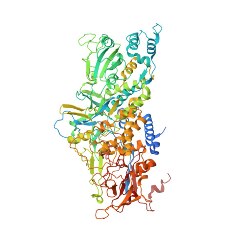
8R0F, 8R0G - PubMed Abstract:
The Giardia lamblia virus (GLV) is a non-enveloped icosahedral dsRNA and endosymbiont virus that infects the zoonotic protozoan parasite Giardia duodenalis (syn. G. lamblia, G. intestinalis), which is a pathogen of mammals, including humans. Elucidating the transmission mechanism of GLV is crucial for gaining an in-depth understanding of the virulence of the virus in G. duodenalis. GLV belongs to the family Totiviridae, which infects yeast and protozoa intracellularly; however, it also transmits extracellularly, similar to the phylogenetically, distantly related toti-like viruses that infect multicellular hosts. The GLV capsid structure is extensively involved in the longstanding discussion concerning extracellular transmission in Totiviridae and toti-like viruses. Hence, this study constructed the first high-resolution comparative atomic models of two GLV strains, namely GLV-HP and GLV-CAT, which showed different intracellular localization and virulence phenotypes, using cryogenic electron microscopy single-particle analysis. The atomic models of the GLV capsids presented swapped C-terminal extensions, extra surface loops, and a lack of cap-snatching pockets, similar to those of toti-like viruses. However, their open pores and absence of the extra crown protein resemble those of other yeast and protozoan Totiviridae viruses, demonstrating the essential structures for extracellular cell-to-cell transmission. The structural comparison between GLV-HP and GLV-CAT indicates the first evidence of critical structural motifs for the transmission and virulence of GLV in G. duodenalis.
- The Laboratory of Molecular Biophysics, Department of Cell and Molecular Biology, Uppsala University, Uppsala, Sweden.
Organizational Affiliation: