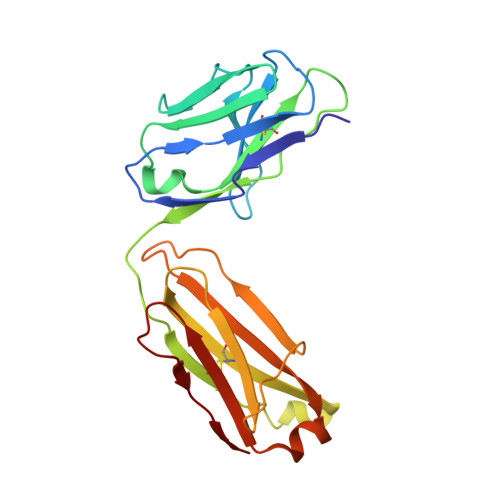
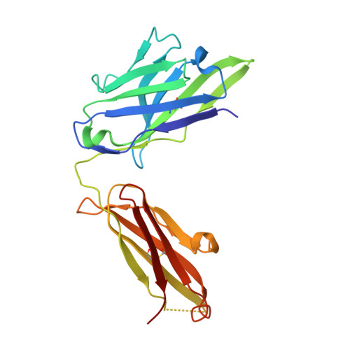
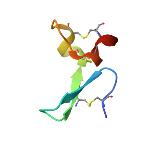
8QY9

J22.9-H, fully humanized Fab Fragment based on chimeric J22.9-xi IgG against BCMA


Experimental Data Snapshot

  • Method: X-RAY DIFFRACTION
  • Resolution: 3.10 Å
  • R-Value Free: 0.301 
  • R-Value Work: 0.254 
  • R-Value Observed: 0.256 

Starting Model: experimental
View more details

wwPDB Validation   3D Report Full Report


This is version 2.2 of the entry. See complete history


Literature

Structure-based humanization of a therapeutic antibody for multiple myeloma.

Marino, S.F.Daumke, O.

(2024) J Mol Med (Berl) 102: 1151-1161

  • DOI: https://doi.org/10.1007/s00109-024-02470-4
  • Primary Citation of Related Structures:  
    8QY9, 8QYA, 8QYB

  • PubMed Abstract: 

    The optimal efficacy of xenogeneically generated proteins intended for application in humans requires that their own antigenicity be minimized. This necessary adaptation of antibodies to a humanized version poses challenges since modifications even distant from the binding sites can greatly influence antigen recognition and this is the primary feature that must be maintained during all modifications. Current strategies often rely on grafting and/or randomization/selection to arrive at a humanized variant retaining the binding properties of the original molecule. However, in terms of speed and efficiency, rationally directed approaches can be superior, provided the requisite structural information is available. We present here a humanization procedure based on the high-resolution X-ray structure of a chimaeric IgG against a marker for multiple myeloma. Based on in silico modelling of humanizing amino acid substitutions identified from sequence alignments, we devised a straightforward cloning procedure to rapidly evaluate the proposed sequence changes. Careful inspection of the structure allowed the identification of a potentially problematic amino acid change that indeed disrupted antigen binding. Subsequent optimization of the antigen binding loop sequences resulted in substantial recovery of binding affinity lost in the completely humanized antibody. X-ray structures of the humanized and optimized variants demonstrate that the antigen binding mode is preserved, with surprisingly few direct contacts to antibody atoms. These results underline the importance of structural information for the efficient optimization of protein therapeutics. KEY MESSAGES: Structure-based humanization of an IgG against BCMA, a marker for Multiple Myeloma. Identification of problematic mutations and unexpected modification sites. Structures of the modified IgG-antigen complexes verified predictions. Provision of humanized high-affinity IgGs against BCMA for therapeutic applications.


  • Organizational Affiliation

    Max Delbrück Centrum for Molecular Medicine, Robert-Rössle-Straße 10, 13125, Berlin, Germany. Stephen-Francis.Marino@bfr.bund.de.


Macromolecules
Find similar proteins by:  (by identity cutoff)  |  3D Structure
Entity ID: 1
MoleculeChains Sequence LengthOrganismDetailsImage
Fab light chain of J22.9-HA [auth L]211Mus musculusMutation(s): 0 
Entity Groups  
Sequence Clusters30% Identity50% Identity70% Identity90% Identity95% Identity100% Identity
Sequence Annotations
Expand
  • Reference Sequence
Find similar proteins by:  (by identity cutoff)  |  3D Structure
Entity ID: 2
MoleculeChains Sequence LengthOrganismDetailsImage
Fab heavy chain of J22.9-HB [auth H]221Mus musculusMutation(s): 0 
Entity Groups  
Sequence Clusters30% Identity50% Identity70% Identity90% Identity95% Identity100% Identity
Sequence Annotations
Expand
  • Reference Sequence
Find similar proteins by:  (by identity cutoff)  |  3D Structure
Entity ID: 3
MoleculeChains Sequence LengthOrganismDetailsImage
Tumor necrosis factor receptor superfamily member 17C [auth A]35Homo sapiensMutation(s): 5 
Gene Names: TNFRSF17BCMBCMA
UniProt & NIH Common Fund Data Resources
Find proteins for Q02223 (Homo sapiens)
Explore Q02223 
Go to UniProtKB:  Q02223
PHAROS:  Q02223
GTEx:  ENSG00000048462 
Entity Groups  
Sequence Clusters30% Identity50% Identity70% Identity90% Identity95% Identity100% Identity
UniProt GroupQ02223
Sequence Annotations
Expand
  • Reference Sequence
Experimental Data & Validation

Experimental Data

  • Method: X-RAY DIFFRACTION
  • Resolution: 3.10 Å
  • R-Value Free: 0.301 
  • R-Value Work: 0.254 
  • R-Value Observed: 0.256 
  • Space Group: C 1 2 1
Unit Cell:
Length ( Å )Angle ( ˚ )
a = 137.36α = 90
b = 55.18β = 121.023
c = 81.16γ = 90
Software Package:
Software NamePurpose
PHENIXrefinement
XDSdata reduction
XDSdata scaling
PHASERphasing

Structure Validation

View Full Validation Report


Entry History & Funding Information

Deposition Data


Funding OrganizationLocationGrant Number
Not funded--

Revision History  (Full details and data files)

  • Version 1.0: 2024-01-17
    Type: Initial release
  • Version 2.0: 2024-07-24
    Type: Coordinate replacement
    Reason: Real space R-factor
    Changes: Advisory, Atomic model, Data collection, Database references, Derived calculations, Non-polymer description, Polymer sequence, Refinement description, Source and taxonomy, Structure summary
  • Version 2.1: 2024-08-14
    Changes: Database references
  • Version 2.2: 2024-09-11
    Changes: Database references