Structure-function characterization of two enzymes from novel subfamilies of manganese peroxidases secreted by the lignocellulose-degrading Agaricales fungi Agrocybe pediades and Cyathus striatus.
Sanchez-Ruiz, M.I., Santillana, E., Linde, D., Romero, A., Martinez, A.T., Ruiz-Duenas, F.J.(2024) Biotechnol Biofuels Bioprod 17: 74-74
- PubMed: 38824538 Search on PubMedSearch on PubMed Central
- DOI: https://doi.org/10.1186/s13068-024-02517-1
- Primary Citation Related Structures:
8QWT, 8QWX, 8QX0 - PubMed Abstract:
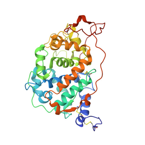
Manganese peroxidases (MnPs) are, together with lignin peroxidases and versatile peroxidases, key elements of the enzymatic machineries secreted by white-rot fungi to degrade lignin, thus providing access to cellulose and hemicellulose in plant cell walls. A recent genomic analysis of 52 Agaricomycetes species revealed the existence of novel MnP subfamilies differing in the amino-acid residues that constitute the manganese oxidation site. Following this in silico analysis, a comprehensive structure-function study is needed to understand how these enzymes work and contribute to transform the lignin macromolecule. Two MnPs belonging to the subfamilies recently classified as MnP-DGD and MnP-ESD-referred to as Ape-MnP1 and Cst-MnP1, respectively-were identified as the primary peroxidases secreted by the Agaricales species Agrocybe pediades and Cyathus striatus when growing on lignocellulosic substrates. Following heterologous expression and in vitro activation, their biochemical characterization confirmed that these enzymes are active MnPs. However, crystal structure and mutagenesis studies revealed manganese coordination spheres different from those expected after their initial classification. Specifically, a glutamine residue (Gln333) in the C-terminal tail of Ape-MnP1 was found to be involved in manganese binding, along with Asp35 and Asp177, while Cst-MnP1 counts only two amino acids (Glu36 and Asp176), instead of three, to function as a MnP. These findings led to the renaming of these subfamilies as MnP-DDQ and MnP-ED and to re-evaluate their evolutionary origin. Both enzymes were also able to directly oxidize lignin-derived phenolic compounds, as seen for other short MnPs. Importantly, size-exclusion chromatography analyses showed that both enzymes cause changes in polymeric lignin in the presence of manganese, suggesting their relevance in lignocellulose transformation. Understanding the mechanisms used by basidiomycetes to degrade lignin is of particular relevance to comprehend carbon cycle in nature and to design biotechnological tools for the industrial use of plant biomass. Here, we provide the first structure-function characterization of two novel MnP subfamilies present in Agaricales mushrooms, elucidating the main residues involved in catalysis and demonstrating their ability to modify the lignin macromolecule.
- Centro de Investigaciones Biológicas Margarita Salas (CIB), CSIC, Ramiro de Maeztu 9, 28040, Madrid, Spain.
Organizational Affiliation: