8QW0

Human NDPK-C in complex with GDP


Experimental Data Snapshot

  • Method: X-RAY DIFFRACTION
  • Resolution: 2.17 Å
  • R-Value Free: 0.229 
  • R-Value Work: 0.189 

Starting Model: experimental
View more details

wwPDB Validation   3D Report Full Report


Ligand Structure Quality Assessment 


This is version 1.1 of the entry. See complete history


Literature

Mechanistic Insights into Substrate Recognition of Human Nucleoside Diphosphate Kinase C Based on Nucleotide-Induced Structural Changes.

Amjadi, R.Werten, S.Lomada, S.K.Baldin, C.Scheffzek, K.Dunzendorfer-Matt, T.Wieland, T.

(2024) Int J Mol Sci 25

  • DOI: https://doi.org/10.3390/ijms25189768
  • Primary Citation of Related Structures:  
    8QVY, 8QVZ, 8QW0, 8QW1, 8QW2, 8QW3

  • PubMed Abstract: 

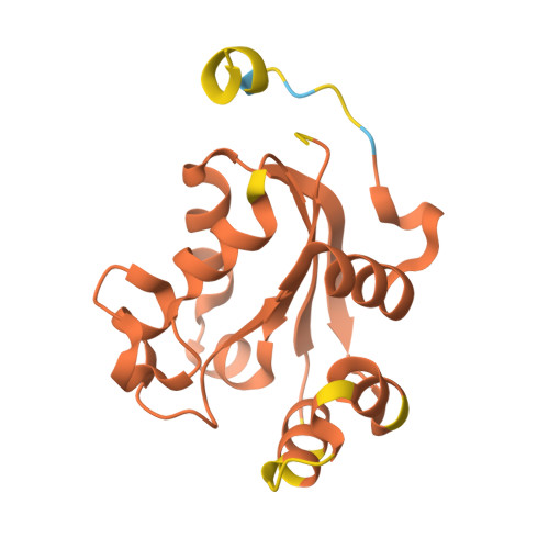
    Nucleoside diphosphate kinases (NDPKs) are encoded by nme genes and exist in various isoforms. Based on interactions with other proteins, they are involved in signal transduction, development and pathological processes such as tumorigenesis, metastasis and heart failure. In this study, we report a 1.25 Å resolution structure of human homohexameric NDPK-C bound to ADP and describe the yet unknown complexes formed with GDP, UDP and cAMP, all obtained at a high resolution via X-ray crystallography. Each nucleotide represents a distinct group of mono- or diphosphate purine or pyrimidine bases. We analyzed different NDPK-C nucleotide complexes in the presence and absence of Mg 2+ and explain how this ion plays an essential role in NDPKs' phosphotransferase activity. By analyzing a nucleotide-depleted NDPK-C structure, we detected conformational changes upon substrate binding and identify flexible regions in the substrate binding site. A comparison of NDPK-C with other human isoforms revealed a strong similarity in the overall composition with regard to the 3D structure, but significant differences in the charge and hydrophobicity of the isoforms' surfaces. This may play a role in isoform-specific NDPK interactions with ligands and/or important complex partners like other NDPK isoforms, as well as monomeric and heterotrimeric G proteins. Considering the recently discovered role of NDPK-C in different pathologies, these high-resolution structures thus might provide a basis for interaction studies with other proteins or small ligands, like activators or inhibitors.


  • Organizational Affiliation

    Institute of Molecular Biochemistry, Medical University of Innsbruck, Innrain 80/82, 6020 Innsbruck, Austria.


Macromolecules
Find similar proteins by:  (by identity cutoff)  |  3D Structure
Entity ID: 1
MoleculeChains Sequence LengthOrganismDetailsImage
Nucleoside diphosphate kinase 3
A, B, C, D, E
A, B, C, D, E, F, G, H, I, J, K, L
155Homo sapiensMutation(s): 0 
Gene Names: NME3
UniProt & NIH Common Fund Data Resources
Find proteins for Q13232 (Homo sapiens)
Explore Q13232 
Go to UniProtKB:  Q13232
PHAROS:  Q13232
GTEx:  ENSG00000103024 
Entity Groups  
Sequence Clusters30% Identity50% Identity70% Identity90% Identity95% Identity100% Identity
UniProt GroupQ13232
Sequence Annotations
Expand
  • Reference Sequence
Small Molecules
Ligands 2 Unique
IDChains Name / Formula / InChI Key2D Diagram3D Interactions
GDP (Subject of Investigation/LOI)
Query on GDP

Download Ideal Coordinates CCD File 
M [auth A]
N [auth B]
O [auth C]
Q [auth D]
S [auth E]
M [auth A],
N [auth B],
O [auth C],
Q [auth D],
S [auth E],
T [auth F],
U [auth G],
V [auth H],
W [auth I],
X [auth J],
Y [auth K],
Z [auth L]
GUANOSINE-5'-DIPHOSPHATE
C10 H15 N5 O11 P2
QGWNDRXFNXRZMB-UUOKFMHZSA-N
SO4
Query on SO4

Download Ideal Coordinates CCD File 
P [auth C],
R [auth D]
SULFATE ION
O4 S
QAOWNCQODCNURD-UHFFFAOYSA-L
Experimental Data & Validation

Experimental Data

  • Method: X-RAY DIFFRACTION
  • Resolution: 2.17 Å
  • R-Value Free: 0.229 
  • R-Value Work: 0.189 
  • Space Group: P 1 21 1
Unit Cell:
Length ( Å )Angle ( ˚ )
a = 104.34α = 90
b = 78.64β = 97.561
c = 112.37γ = 90
Software Package:
Software NamePurpose
REFMACrefinement
XDSdata reduction
XSCALEdata scaling
PHASERphasing

Structure Validation

View Full Validation Report


Ligand Structure Quality Assessment 


Entry History & Funding Information

Deposition Data


Funding OrganizationLocationGrant Number
Not funded--

Revision History  (Full details and data files)

  • Version 1.0: 2024-09-18
    Type: Initial release
  • Version 1.1: 2024-10-09
    Changes: Database references, Structure summary