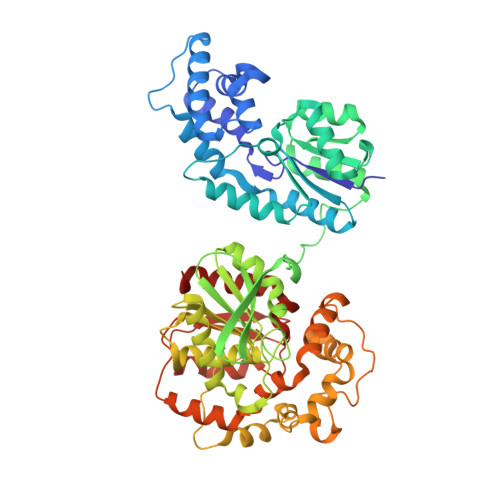
8QVM

Comparison of room-temperature and cryogenic structures of soluble Epoxide Hydrolase with ligands bound.


Experimental Data Snapshot

  • Method: X-RAY DIFFRACTION
  • Resolution: 2.00 Å
  • R-Value Free: 0.246 
  • R-Value Work: 0.197 
  • R-Value Observed: 0.199 

Starting Model: experimental
View more details

wwPDB Validation   3D Report Full Report


This is version 1.1 of the entry. See complete history


Literature

Exploring serial crystallography for drug discovery.

Dunge, A.Phan, C.Uwangue, O.Bjelcic, M.Gunnarsson, J.Wehlander, G.Kack, H.Branden, G.

(2024) IUCrJ 11: 831-842

  • DOI: https://doi.org/10.1107/S2052252524006134
  • Primary Citation of Related Structures:  
    8QVF, 8QVG, 8QVH, 8QVK, 8QVL, 8QVM, 8QWG, 8QWI

  • PubMed Abstract: 

    Structure-based drug design is highly dependent on the availability of structures of the protein of interest in complex with lead compounds. Ideally, this information can be used to guide the chemical optimization of a compound into a pharmaceutical drug candidate. A limitation of the main structural method used today - conventional X-ray crystallography - is that it only provides structural information about the protein complex in its frozen state. Serial crystallography is a relatively new approach that offers the possibility to study protein structures at room temperature (RT). Here, we explore the use of serial crystallography to determine the structures of the pharmaceutical target, soluble epoxide hydrolase. We introduce a new method to screen for optimal microcrystallization conditions suitable for use in serial crystallography and present a number of RT ligand-bound structures of our target protein. From a comparison between the RT structural data and previously published cryo-temperature structures, we describe an example of a temperature-dependent difference in the ligand-binding mode and observe that flexible loops are better resolved at RT. Finally, we discuss the current limitations and potential future advances of serial crystallography for use within pharmaceutical drug discovery.


  • Organizational Affiliation

    Department of Chemistry and Molecular Biology, University of Gothenburg, Box 462, SE-405 30 Gothenburg, Sweden.


Macromolecules
Find similar proteins by:  (by identity cutoff)  |  3D Structure
Entity ID: 1
MoleculeChains Sequence LengthOrganismDetailsImage
Bifunctional epoxide hydrolase 2549Homo sapiensMutation(s): 0 
Gene Names: EPHX2
UniProt & NIH Common Fund Data Resources
Find proteins for P34913 (Homo sapiens)
Explore P34913 
Go to UniProtKB:  P34913
PHAROS:  P34913
GTEx:  ENSG00000120915 
Entity Groups  
Sequence Clusters30% Identity50% Identity70% Identity90% Identity95% Identity100% Identity
UniProt GroupP34913
Sequence Annotations
Expand
  • Reference Sequence
Small Molecules
Ligands 1 Unique
IDChains Name / Formula / InChI Key2D Diagram3D Interactions
PGE
Query on PGE

Download Ideal Coordinates CCD File 
B [auth A]TRIETHYLENE GLYCOL
C6 H14 O4
ZIBGPFATKBEMQZ-UHFFFAOYSA-N
Experimental Data & Validation

Experimental Data

  • Method: X-RAY DIFFRACTION
  • Resolution: 2.00 Å
  • R-Value Free: 0.246 
  • R-Value Work: 0.197 
  • R-Value Observed: 0.199 
  • Space Group: P 65 2 2
Unit Cell:
Length ( Å )Angle ( ˚ )
a = 94.41α = 90
b = 94.41β = 90
c = 247.57γ = 120
Software Package:
Software NamePurpose
BUSTERrefinement
CrystFELdata reduction
CrystFELdata scaling
PHASERphasing

Structure Validation

View Full Validation Report


Entry History & Funding Information

Deposition Data


Funding OrganizationLocationGrant Number
The Swedish Foundation for Strategic ResearchSwedenID17-0060
Swedish Research CouncilSweden2017-06734
Swedish Research CouncilSweden2021-05662
Swedish Research CouncilSweden2021-05981

Revision History  (Full details and data files)

  • Version 1.0: 2024-08-14
    Type: Initial release
  • Version 1.1: 2024-09-11
    Changes: Database references