Download Mendeley
A strategy to design protein-based antagonists against type I cytokine receptors.
Ullrich, T., Klimenkova, O., Pollmann, C., Lasram, A., Hatskovska, V., Maksymenko, K., Milijas-Jotic, M., Schenk, L., Lengerke, C., Hartmann, M.D., Piehler, J., Skokowa, J., ElGamacy, M.(2024) PLoS Biol 22: e3002883-e3002883
- PubMed: 39591631 Search on PubMedSearch on PubMed Central
- DOI: https://doi.org/10.1371/journal.pbio.3002883
- Primary Citation Related Structures:
8QUP - PubMed Abstract:
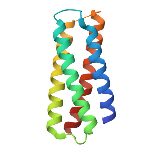
Excessive cytokine signaling resulting from dysregulation of a cytokine or its receptor can be a main driver of cancer, autoimmune, or hematopoietic disorders. Here, we leverage protein design to create tailored cytokine receptor blockers with idealized properties. Specifically, we aimed to tackle the granulocyte-colony stimulating factor receptor (G-CSFR), a mediator of different types of leukemia and autoinflammatory diseases. By modifying designed G-CSFR binders, we engineered hyper-stable proteins that function as nanomolar signaling antagonists. X-ray crystallography showed atomic-level agreement with the experimental structure of an exemplary design. Furthermore, the most potent design blocks G-CSFR in acute myeloid leukemia cells and primary human hematopoietic stem cells. Thus, the resulting designs can be used for inhibiting or homing to G-CSFR-expressing cells. Our results also demonstrate that similarly designed cytokine mimics can be used to derive antagonists to tackle other type I cytokine receptors.
- Max Planck Institute for Biology, Department of Protein Evolution, Tübingen, Germany.
Organizational Affiliation: